
Figure 4.
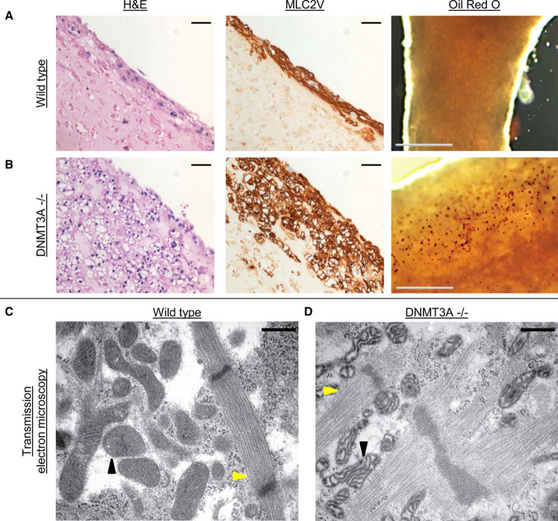
Morphology of DNA methyltransferase(DNMT) 3A knockout engineered heart tissue(EHT).
Histological analysis of (A) wild-type and (B) DNMT3A−/− EHTs. Left, Hematoxylin and eosin staining (H&E); scale bar, 50 µm. Middle, MLC2V staining; scale bar, 50 µm. Right, Lipid staining with Oil Red O; scale bar, 500 µm, red dots mark lipid accumulations. Transmission electron microscopy of wild-type (C) and knockout (D) EHTs showing intact sarcomeric structures but degenerated mitochondria in knockout EHT. Yellow arrowheads, sarcomeres; black arrowheads, mitochondria. Scale bar, 500 nm.