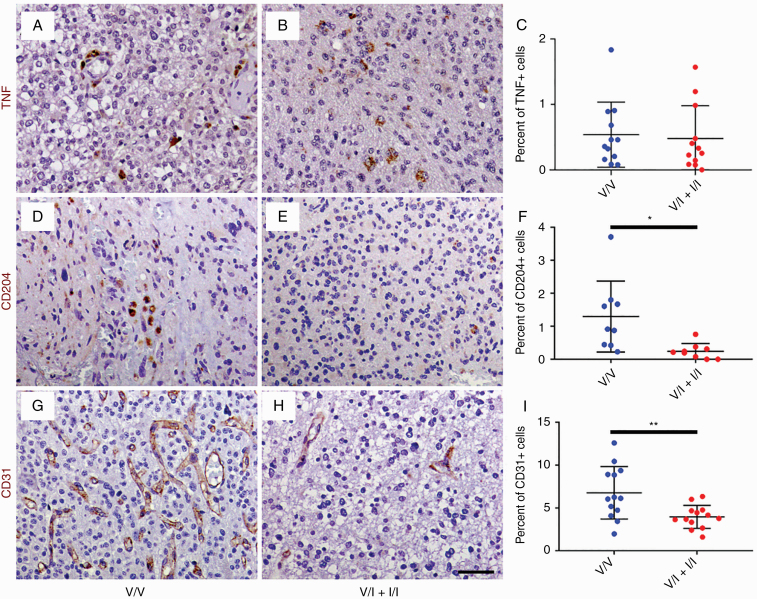
Fig. 3.
Histological sections of WHO grade III gliomas from V/V (A, D, and G) and V/I + I/I patients (B, E, and H) were immunostained with antibodies against M1 macrophage marker TNF (brown, A and B), M2 macrophage marker CD204 (brown, D and E), and endothelial marker CD31 (brown, G and H), followed by counterstaining with hematoxylin (blue; A, B, D, E, G, and H). There were no significant differences in density of TNF-positive cells in HGGs from V/V (n = 12) and V/I patients (n = 12; C). On the other hand, HGGs from V/V patients (n = 9) demonstrated significantly increased density of CD204-positive macrophages compared with HGGs from V/I + I/I patients (n = 10; F). Similarly, V/V tumors (n = 13) showed significantly increased density of CD31-positive endothelial cells compared with V/I + I/I tumors (n = 13; I). *P < 0.05, **P < 0.01, unpaired t-test. Scale bar, 50 μm.

