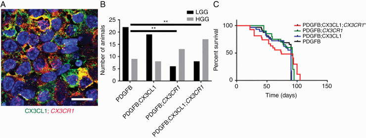
Fig. 4.
(A) Histological sections of murine gliomas induced by RCAS-PDGFB together with RCAS-CX3CL1 and RCAS-CX3CR1 were immunostained with antibodies against CX3CL1 (green) and CX3CR1 (red), followed by counterstaining with DAPI (blue), revealing membranous coexpression of CX3CL1 and CX3CR1. (B) Significantly higher proportion of HGGs were seen in mice injected with RCAS-PDGFB and RCAS-CX3CR1 (13/19) as well as in mice injected with RCAS-PDGFB, RCAS-CX3CL1, and RCAS-CX3CR1 (17/25) compared with mice injected with RCAS-PDGFB and RCAS-CX3CL1 (8/27) or with RCAS-PDGFB alone (9/31; **P < 0.01, chi-squared test). (C) Kaplan–Meier survival curves for PDGFB (n = 31), PDGFB;CX3CL1 (n = 29), PDGFB;CX3CR1 (n = 23), and PDGFB;CX3CL1;CX3CR1 (n = 28) groups show significantly shorter OS for PDGFB;CX3CL1;CX3CR1 group compared with PDGFB group (*P < 0.05, log-rank test). Scale bar, 25 μm.

