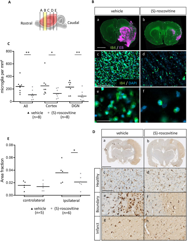
Fig. 4.
(S)-roscovitine decreases microglia’s number. (A) Rostro-caudal position of slices B, used for analysis. (B) Immunohistofluorescent analysis on slice B of EB and microglia (a, b) in (S)-roscovitine (n = 8) and vehicle group (n = 8) (Scale bar length is 2 mm). Isolectin B4 marking was used for microglia count (c, d) (Scale bar length is 100 µm). Microglia were differentiated from blood vessels by morphological observation (e, f and Supplementary Fig. 3) (Scale bar length is 50 µm). Measurements were performed by blind operator. Number of microglia was measured on cortex and DGN and combined in total measurement (C). (D) Immunohistochemical analysis of the second experimental protocol on paraffin-embedded tissue of (S)-roscovitine treated animal (n = 6) and vehicle-treated animal (n = 5). Microglia were labelled with IBA1 antibody coupled with horseradish peroxidase and stained by diaminobenzidine. Microglia density was measured on healthy cH (c, d), on boundary (e, f) and infarct (g, h) region of cortex. IBA1-positive area fraction in border and infarct were combined to calculate ipsilateral area fraction (E). ROI were placed by blind operator and area fraction calculation was performed with NIS-Elements program (Nikon). a, b: Scale bars length is 2 mm; c–h: Scale bars length is 100 µm. * P ≤ 0.05; ** P ≤ 0.01.

