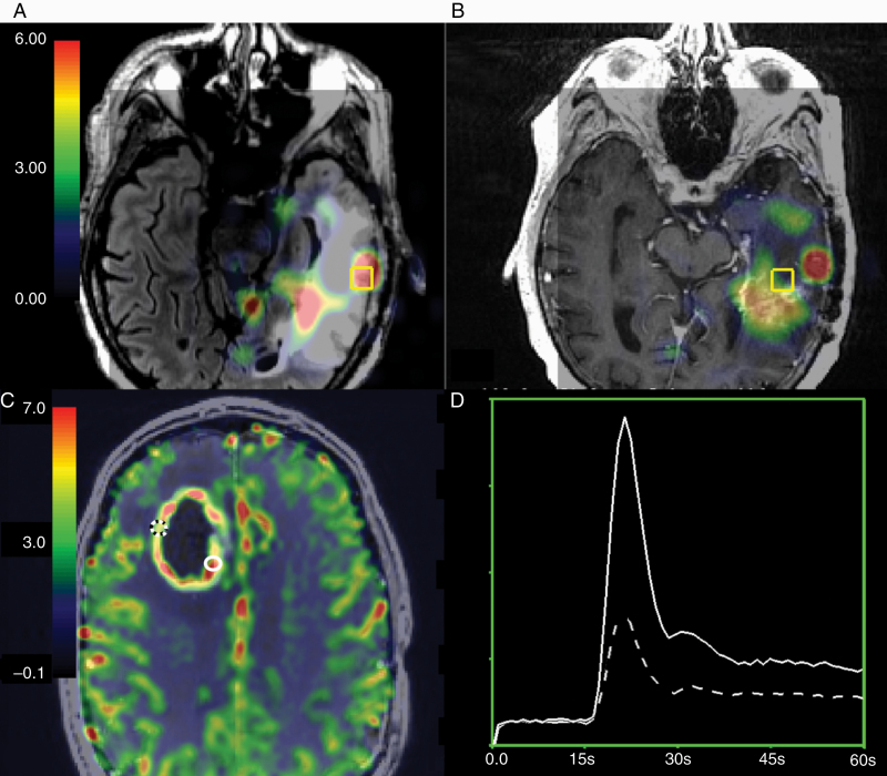
Fig. 2.
Within-patient imaging differences between treatment-induced injury and recurrent HGG. (A) Non-enhancing CNI parameter maps overlaid on the T2 FLAIR image are elevated in HGG samples. (B) Reduced CNI in a sample with treatment-induced injury within the same patient (overlaid on the post-contrast T1-weighted image). (C) Enhancing rCBV parameter maps are elevated in HGG (solid white location) versus treatment-induced injury (dashed location). (D) The corresponding ΔR2* curves.

