Figure 3.

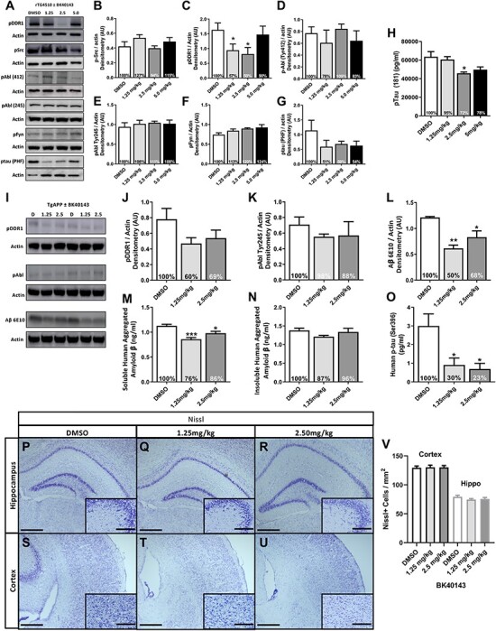
BK40143 specifically inhibits DDR1 and lowers toxic proteins in AD models. (A) Representative western blots of pDDR1, pSrc, pAbl (Tyr412 and Tyr 245), pFyn and pTau paired-helical filament (PHF) from 3 to 4 months old rTG4510 mice treated with 1.25, 2.5 and 5 mg/kg BK40143 or DMSO for 7 days and quantification of (B) pDDR1, (C) pSrc, (D) pAbl (Tyr412), (E) pAbl (Tyr245), (F) pFyn and (G) p-tau (PHF), each normalized to actin with percent change from DMSO-treated animals. (H) ELISA for human pTau (Thr181) (pg/ml) from whole brain. *P < 0.05; normal one-way ANOVA; n = 4 per treatment group. (I–V) Western blot, ELISA and Nissl staining from 7 to 8 months old TgAPP mice treated with 1.25 and 2.5 mg/kg of BK40143 or DMSO for 21 days. (I) Representative western blots of pDDR1, pAbl and Aβ (6E10) with corresponding actin. Quantification of (J) pDDR1 (Tyr796), (K) pAbl (Tyr254) and (L) Aβ (6E10), each normalized to actin with percent change from DMSO-treated group. *P < 0.05, **P < 0.01; normal one-way ANOVA or Student’s t-test. n = 5 per treatment group. ELISA for (M) intracellular soluble human aggregated Aβ (ng/ml), (N) extracellular insoluble human aggregated Aβ (ng/ml) and (O) human p-tau (Ser396) from whole brain. *P < 0.05 and ***P < 0.001; normal one-way ANOVA; n = 5 per treatment group. (P–V) Nissl staining and quantification of the hippocampus and cortex. (P–R) Representative images of hippocampal Nissl staining in (P) DMSO, (Q) 1.25 and (R) 2.5 mg/kg BK40143-treated TgAPP mice. Scale bars, 500 and 20 μm inlay. (S–U) Representative images of cortical Nissl staining in (S) DMSO, (T) 1.25 and (U) 2.5 mg/kg BK40143-treated TgAPP mice. (V) Quantification of Nissl positive cells in the cortex and hippocampus, n = 5 per group, n = 4 coronal sections per mouse and three images per section for quantification.
