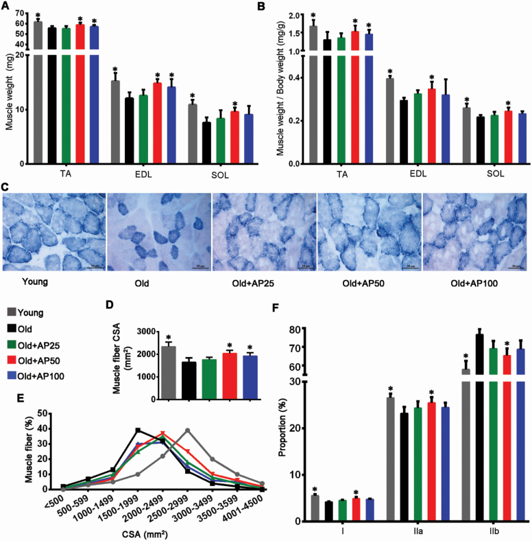
Figure 2.
Apigenin attenuated muscle atrophy in aged mice. (A) Weights of tibialis anterior (TA), extensor digitorum longus (EDL), and soleus (SOL) muscles were normalized to tibia length. (B) Relative weights of TA, EDL, and SOL normalized to body weight. (C) Succinate dehydrogenase (SDH) staining was performed on 10-μm-thick sections from TA muscles frozen in liquid nitrogen-chilled isopentane. Scale bar: 50 μm. (D) Average fiber size (cross-sectional area, CSA) of the SDH-stained TA muscles. (E) Muscle fiber frequency distribution of the SDH-stained TA muscle. (F) Numbers of types I (slow oxidative), IIa (fast oxidative glycolytic), and IIb (fast twitch glycolytic) muscle fibers. Data are presented as the mean ± SD (n = 8–10). *p < .05, versus the Old group (25 months).

