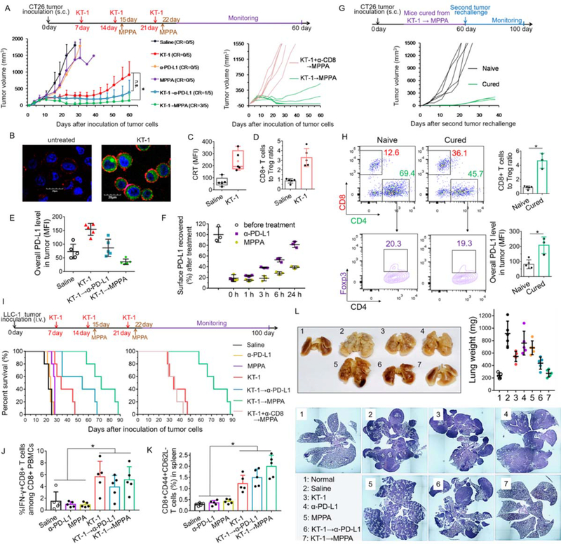
Figure. 6. Anti-tumor and anti-metastatic effects of KT-1 and MPPA combination in subcutaneous CT26 and metastatic LLC-1 tumor models.
(A) CT26 colon tumor growth curves after indicated treatments (n=5). BALB/c mice were subcutaneously inoculated with 2×10⁶ CT26 cells on day 0. On days 7, 14, and 21, tumor-bearing mice were treated with KT-1. On days 15, and 22, mice were treated with anti-PD-L1 therapy, α-PD-L1 antibodies or MPPA conjugates. CD8-depleting antibodies were given simultaneously with KT-1 to mice subjected to CD8+ T-cell ablation. The arrows indicate the treatment regimens for KT-1 and MPPA combination. (B) Confocal images of KT-1-enhanced CRT exposure on the surface of CT26 cells in vitro. Blue: cell nuclei; Green: EPI; Red: CRT. In vivo (C) CRT up-regulation on cell surface, and (D) CD8+ T cells to Treg ratio, after two doses treatments (on Day 7 and Day 14) with saline and KT-1 for CT26 tumor-bearing mice. (E) In vivo PD-L1 expressions in CT26 tumors (on Day 17) after two doses treatment (on Day 7 and Day 14) with KT-1, followed by one dose (Day 15) treatment with α-PD-L1 or MPPA. (F) Time-dependent recovery of surface PD-L1. CT26 tumor cells were isolated from tumor-bearing mice after two doses treatment (on Day 7 and Day 14) with KT-1. Then cell surface was precoated with saturating concentration of α-PD-L1 or MPPA at 4 °C for 2 h. Afterward, cells were washed and incubated with fresh culture medium at 37 °C. At selected time points (0, 1, 3, 6, 24 h), surface accessible PD-L1 receptors were stained with fluorophore-labeled anti-PD-L1 antibody and measured by flow cytometry. (G) Individual tumor volume measurement after naive control mice (n=5) or KT-1→MPPA treated CR mice (n=3) in (A) were subcutaneously re-challenged with CT26 cells. (H) Immune status including CD8+ T cells, Tregs, PD-L1 expression in primary CT26 tumors of naive mice and secondary CT26 tumors of cured mice. (I) Survival rate of mice after indicated treatments (n=5). C57BL/6 mice were intravenously inoculated with 2×10⁵ LLC-1 Lewis lung carcinoma cells on day 0. Then mice were treated as described in (A). (J) Percentage of tumor cell-reactive T cells (IFN-γ+CD8+) among PBMCs against LLC-1 cells, (K) CD44+CD62L- memory effector CD8+ T cells in spleen, and (L) Tumor burden in lungs depicted as lung weight and hematoxylin-eosin histology analysis of lung lobe sections, from mice in (I) on Day 25 or their endpoint,. *P < 0.05, n.s, not significant, one-way ANOVA with Tukey’s multiple comparison test.

