
Figure 2. UPF2 is required for surface expression of GLUR1 on dendrites.
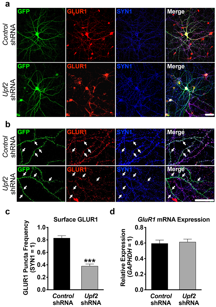
a-b, Hippocampal neurons were infected at DIV7 with control- or Upf2-shRNA lentivirus and fixed at DIV21. Surface GLUR1 expression was detected using an anti-N-terminus-GluR1 antibody (see Methods). Arrows indicate positive and negative GLUR1 signal at spines.
c, UPF2 positively regulates surface GLUR1 levels.
Quantifications of surface GLUR1 frequency in dendrites (n=3/group; 10 neurons-24 dendrites/group). Knockdown of Upf2 did not change SYN1 density (see Figure S4 for PSD-95 staining) indicating unaltered synaptic potential.
d, Knockdown of Upf2 does not alter GluR1 mRNA levels.
qRT-PCR of GluR1 mRNA levels in control- and Upf2-shRNA infected neurons (n=3/group; see Figure S7 for infection efficiency).
Data are represented as mean ± (SEM); ***p < 0.001. Scale bar: 20 μm.