Fig. 4.
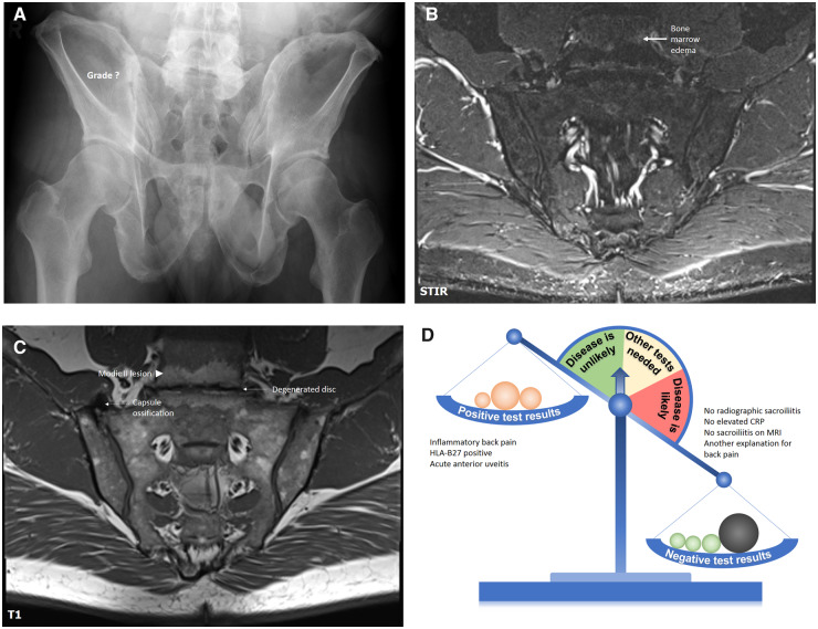
Conventional radiography, MRI-STIR, MRI-T1 of sacroiliac joints, and a diagnostic scale for patient B
A 53-year-old male patient referred by an ophthalmologist because of two recent episodes of acute anterior uveitis and inflammatory back pain of intermittent intensity for about 20 years. HLA-B27 is positive, CRP is normal. Conventional radiography of sacroiliac joints showed suspicious changes, but no SpA-compatible changes could be found on MRI. At the same time, degenerative changes of the intervertebral disc represent the most likely explanation of back pain in this case. See the article text for further details. SpA: spondyloarthritis; STIR: short tau inversion recovery.

