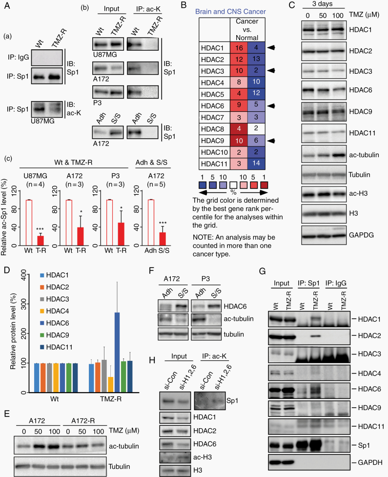
Fig. 1.
Sp1 is deacetylated by HDAC1/2/6 in TMZ-resistant GBM cells. (A) The wild-type (Wt) and TMZ-resistant (TMZ-R) GBM cells,12 as well as GBM spheroids formed in serum-free medium/suspension (S/S) culture and the control attached (Adh) cells13 were used for the immunoprecipitation (IP) assay with rabbit IgG, anti-Sp1 (Sp1, panel a), and anti–acetyl-lysine (ac-K, panels b and c) antibodies, and analyzed using immunoblotting (IB) as indicated. In panel c, the protein level of acetylated Sp1 was normalized to its total protein and quantified. (B) Gene expression profiles of HDACs in brain tumors were analyzed using the Oncomine database. HDAC1/3/6/9, shown by the arrows, were upregulated more in cancer tissues than in normal samples. Red indicates upregulation; blue indicates downregulation. The number in the cell represents the number of datasets that pass the filter criteria (threshold: P < 0.05). (C to F) Cells were harvested and analyzed using IB. The Wt (C and E) and TMZ-R (E) A172 cells were treated with the indicated concentrations of TMZ for 3 days. (D) The protein expression of HDACs in Wt and TMZ-R P11 GBM cells was normalized to the loading control and quantified. (F) The levels of HDAC6 and tubulin acetylation in attached GBM cells and tumorspheres. (G) Wt and TMZ-R P11 cells were used for IP assay with anti-Sp1 antibodies and rabbit IgG, and analyzed using IB as indicated. (H) TMZ-R U87MG cells were transfected with a nontargeting control siRNA or HDAC1/2/6-specific siRNAs as indicated. After knockdown, the cells were used for IP assay. (t-test: *P < 0.05, ***P < 0.001)

