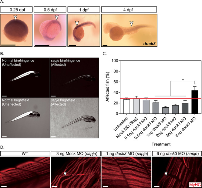
Figure 2.

Knockdown of dock3 mRNA in DMD zebrafish reduces dystrophic pathology. (A) ISH of zebrafish dock3 mRNA during early developmental time points. Note the robust expression of zebrafish dock3 mRNA in muscle tissues at stages of early muscle formation and muscle pioneer cell fusion. Not shown sense probes are used as internal controls. Arrowheads demarcate dock3 mRNA ISH signal. Scale bar = 100 μm. (B) Representative images of normal and sapje mutant birefringence morphology. Scale bar = 200 μm. (C) Quantification of muscle birefringence shows that 1 ng of dock3 MO improves muscle birefringence scoring in sapje mutant zebrafish, while 6 ng of dock3 MO worsens muscle birefringence scoring. (n = 3 experimental replicates; one-way ANOVA with Tukey’s correction; *P < 0.05). (D) MyHC (F-59 antibody) immunofluorescent staining of 4 dpf WT (uninjected) or sapje mutant larvae injected with control (mock) morpholino, or dock3 MO (1 or 6 ng). Arrowheads demarcate myofiber tears from the sarcolemmal membrane. Scale bar = 40 μm.
