Figure 7.

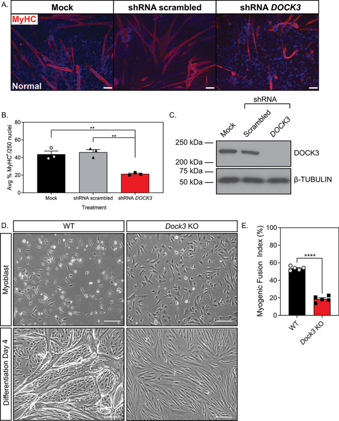
DOCK3 is essential for normal myoblast fusion. (A) MyHC staining reveals that knockdown of DOCK3 protein in normal human primary myotubes decreases myogenic fusion indices. MyHC (MF-20 antibody, DSHB Iowa, red) immunofluorescence of Day 7 differentiated primary myotubes along with DAPI (blue). MOCK, shRNA scrambled and shRNA DOCK3 lentiviral particles were transduced at an MOI of 10. Scale bar = 50 μm. (B) Average myogenic fusion indices as calculated by percentage of nuclei inside of MyHC+ cells out of 250 nuclei as previously described (68). Myotubes with shRNA DOCK3 knockdown have significantly less myogenic fusion than shRNA scrambled control (n = 3 experimental replicates; one-way ANOVA with Tukey’s correction; *P < 0.05). (C) Western blot analysis of whole cell lysates taken from normal human Day 7 differentiated myotubes transduced with mock (control), scrambled (shRNA internal control) or shRNA DOCK3 knockdown inhibitor lentiviral particles showing successful knockdown of DOCK3 protein expression. (D) Representative images of WT and Dock3 KO mouse myoblasts undergoing differentiation. (E) Quantification of myogenic fusion index where Dock3 KO mouse myoblasts has significantly less myogenic fusion than WT mouse myoblasts. Scale bar = 100 μm. (n = 5; student’s t-test, two-tailed; ****P < 0.0001).
