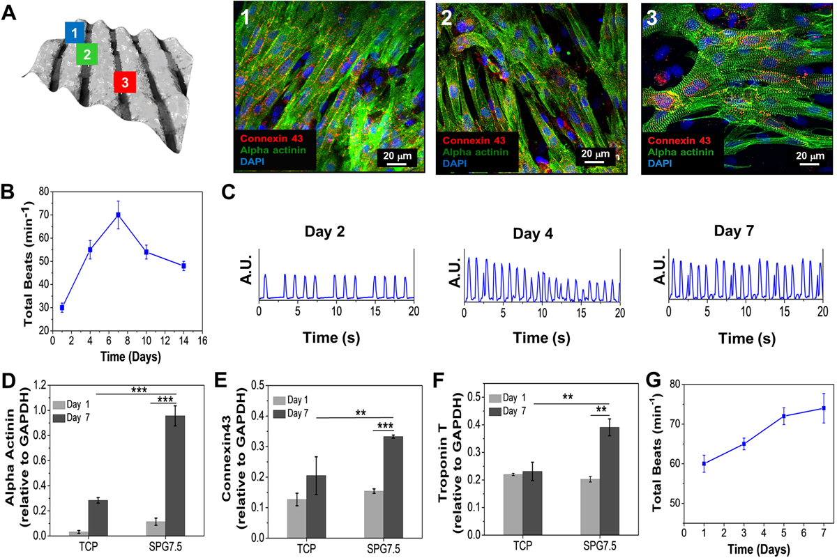
Figure 4.

Functional assessment of cardiomyocytes seeded 3D printed constructs. (A) Computed bright field image of the 3D printed scaffold showing cell attachment on different microfibers; the vertical fibers as (1), horizontal fibers as (2) and the spaces in between them as (3). The seeded NRCMs on the three regions were analyzed for their orientation and maturation via immunostaining. Beating behavior of the seeded (B) NRCMs 3D printed micro-fibrous scaffold (n=5). (C) The beating signal of the seeded NRCMs on day 2, 4, and 7. (D-F) Gene expression analysis of cardiac specific biomarkers expressed by NRCMs seeded on 3D printed constructs (n=3; *** p ≤ 0.001 and **p ≤ 0.01). (G) Beating behavior of the seeded hiPSC-CMs on the 3D printed micro-fibrous scaffold (n=3).
