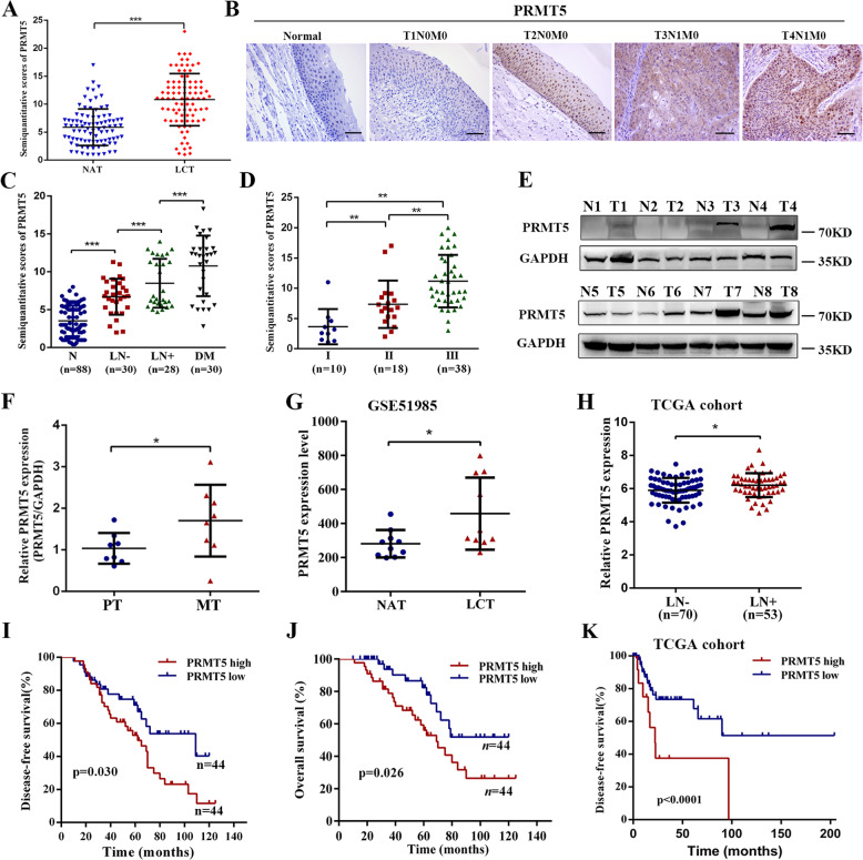
Fig. 1. High PRMT5 expression in laryngeal carcinoma is correlated with lymph-node metastasis and unfavorable prognosis.
a The levels of PRMT5 in 88 paired laryngeal carcinoma tissues (LCT) and normal adjacent tissues (NAT). b Representative IHC staining of PRMT5 expression in paraffin-embedded normal tissues and laryngeal carcinoma with or without lymph-node metastasis. Scale bar, 50 µm. c IHC staining statistical analysis of PRMT5 in normal tissues (N, n = 88), primary LCT without (LN−, n = 30) or with (LN+, n = 28) regional lymph-node and distant metastasis (DM, n = 30). Mean + SD. d IHC staining statistical analysis of the association between PRMT5 expression status and TNM stage. e, f PRMT5 expression in eight paired laryngeal carcinoma lymph-node metastasis (MT) and adjacent primary tissues (PT) was performed by western blotting and qPCR. g PRMT5 expression was detected in LCT and NAT from GSE51986 cohort. h PRMT5 expression was detected in LCT without (LN−, n = 70) or with (LN+, n = 53) lymph-node metastasis in TCGA cohort. i, j Kaplan–Meier analysis for disease-free and overall survival of laryngeal carcinoma patients with high versus low expression of PRMT5 in our cohort. k Kaplan–Meier curves for DFS of laryngeal carcinoma patients with high versus low expression of PRMT5 in TCGA cohort. Statistical significance was assessed using a two-tailed t test. *p < 0.05, **p < 0.01, ***p < 0.001.

