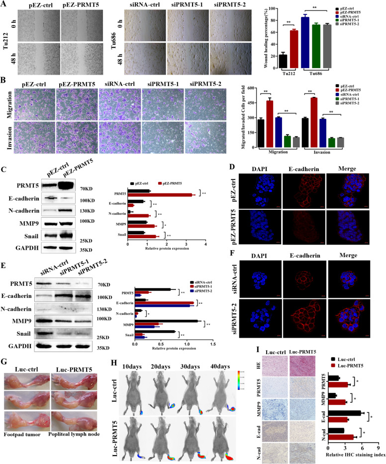
Fig. 3. PRMT5 promotes cancer metastasis in vitro and in vivo.
a Representative images and quantification of wound-healing assays using Tu212 and Tu686 cells showing cell mobility after overexpression or knockdown of PRMT5 (left panels); a histogram analysis of cell migration distances is shown (right panels). Scale bar, 100 µm. b Representative images and quantification of migration and invasion abilities in PRMT5-transduced Tu212, and PRMT5-silenced Tu686 and control cells. Scale bar, 100 µm. c The effect of PRMT5 overexpression on EMT marker expression was assessed by western blotting. d Immunofluorescence images for E-cadherin expression in Tu212 cells. e The effect of PRMT5 knockdown on EMT marker expression in Tu686 cells. f Immunofluorescence images for E-cadherin expression in Tu686 cells. Scale bar, 10 µm. g The nude mouse model of popliteal lymph-node metastasis. Tu212 cells stably transfected with control and PRMT5 were injected into the footpads of the nude mice, and the popliteal lymph nodes were enucleated and analyzed. h Bioluminescent images of primary and metastatic tumors were monitored at 10, 20, 30, and 40 days post treatment. i Representative images of tumor nodes were stained with H&E. The expression of PRMT5, MMP9, E-cadherin, and N-cadherin in the lymph-node metastasis model was detected by immunohistochemistry. Scale bar, 50 µm. All the experiments were repeated three times, and the results are presented as mean ± SD. Statistical significance was assessed using two-tailed t tests. *p < 0.05, **p < 0.01.

