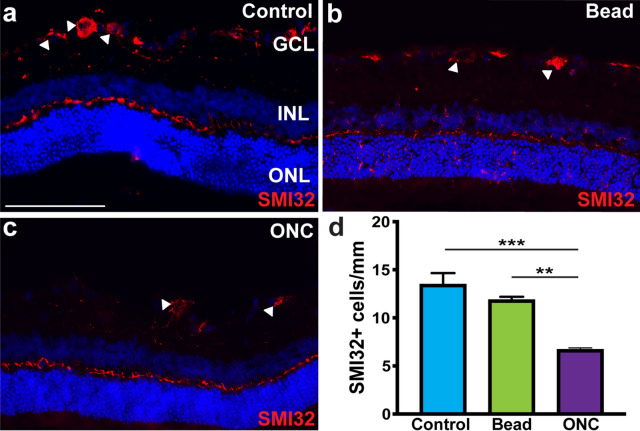
Figure 3.
Alpha-RGCs display susceptibility to injury. (a) Alpha-RGCs were identified within the ganglion cell layer by the expression of SMI32. (b–d) Following ONC, the expression of SMI32 was significantly decreased compared to both controls and the bead occlusion model. Error bars represent S.E.M. Scale bars equal 100 µm. Statistical differences were determined using a One-way ANOVA followed by Tukey’s post hoc with 95% confidence. (**p < 0.01, ***p < 0.001). n = 6 retinas were analyzed per group, with 4 cross sections and numerous technical replicates per cross section. Cross sections contained both nasal and temporal parts, with technical replicates including areas from both the central and peripheral retina.

