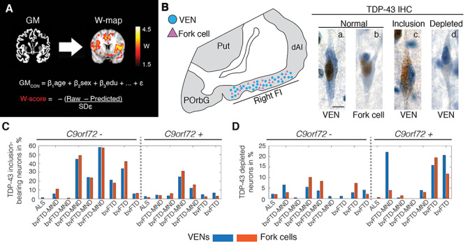
Figure 1.

Patient assessment. (A) All patients underwent structural MRI. Images were preprocessed, segmented into gray matter tissue intensity maps, and transformed to W-score maps using a multiple regression model trained on healthy older adults (GMcon) to model the voxel-wise relationship between relevant demographic variables and segmented tissue intensity. The model provides a predicted gray matter map for each patient, and the actual and predicted gray matter maps are compared to derive W-score maps, which reflect the deviation at each voxel from the predicted gray matter intensity. To facilitate the interpretation of these maps, the sign of W-score values was inverted, with higher W-score values reflecting higher levels of atrophy. (B) Postmortem, the rate of VENs (blue circles) and fork cells (violet triangles) with TDP-43 pathobiology was quantified in layer 5 of the right FI via unbiased counting on TDP-43 immunostained sections. TDP-43 is normally expressed in the nucleus (brown nucleus in [a.] normal VEN and [b.] normal fork cell), which in patients aggregates into neuronal cytoplasmic inclusions ([c.] TDP-43 inclusion-bearing VEN) while being cleared from the nucleus. A minority of neurons lacks either normal nuclear TDP-43 or a cytoplasmic inclusion ([d.] nuclear TDP-43 depleted VEN). Scale bar in B.a. represents 10 μm. Percentage of (C) TDP-43 inclusion-bearing and (D) TDP-43 depleted VENs (blue) and fork cells (orange) for individual patients. ALS = amyotrophic lateral sclerosis; bvFTD = behavioral variant of frontotemporal dementia; bvFTD-MND = behavioral variant of frontotemporal dementia with motor neuron disease; C9orf72— = C9orf72 expansion noncarriers; C9orf72 + = C9orf72 expansion carriers; dAI = dorsal anterior insula; FI = frontoinsular cortex; IHC = immunohistochemistry; GM = gray matter; POrbG = posterior orbitofrontal gyrus; Predicted = through the healthy adults regression model predicted patient’s gray matter map; Put = putamen; Raw = segmented patient’s gray matter map; SDε = standard deviation of the residuals in the healthy adults model; TDP-43 = transactive response DNA binding protein with 43 kD; VEN = von Economo neuron.
