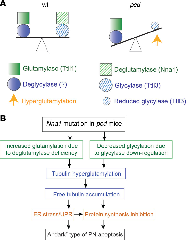
Figure 6. Schematic representation of the molecular mechanisms.
(A) The molecular seesaw model on the left shows the WT balance between glutamylase (Ttll1) and deglutamylase (Nna1) on the seesaw’s left side and between glycylase protein (Ttll3) and a presumed deglycylase or other molecular mechanism on the seesaw’s right side. The molecular seesaw model on the right shows the pcd abnormal unbalance between the increased glycylase (measured in response to the net increase in tubulin that contains polyglutamate) and the decreased glycylation contrasting with the tubulin hyperglutamylation in pcd PNs, beginning at about P20. (B) The pathological pathway through which the Nna1 mutation leads to a dark type of P20 pcd PN death. Ttll1, tubulin tyrosine ligase–like 1; Nna1, neuronal nuclear protein induced by axotomy; PNs, Purkinje neurons; pcd, Purkinje cell degeneration.

