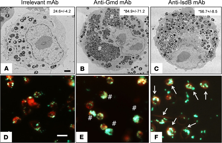
Figure 4. Anti-IsdB mAb induces increased S. aureus internalization by macrophages.
RAW 264.7 cells grown in serum-containing media were challenged with MRSA (USA300LAC) treated with 50 μg/mL(A) irrelevant IgG (negative control), (B) anti-Gmd (1C11 positive control), or (C) anti-IsdB, for 2 hours at (MOI = 10), before TEM as described in Methods. Representative images (original magnification, ×5000) are shown with quantification of the number of bacteria per cell (mean ± SD, n = 4, *P < 0.05 vs. IgG control via Kruskal-Wallis test; scale bar: 2 μm.). This experiment was repeated with LysoTracker Red–labeled RAW cells challenged with GFP+ UAMS-1 via real-time fluorescent microscopy (original magnification, ×100) (D–F). Note the megaclusters (pound signs indicates clustered GFP signal in E) and Trojan horse macrophages (arrows indicate punctate GFP signal in F). Scale bar: 20 μm.

