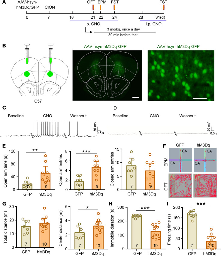
Figure 2. Activation of VLO neurons induced an antianxiodepressive effect in TN mice.
(A) Schematic of the protocol for the experiments in E–I. (B) Schematic and photomicrograph of coronal section showing AAV-hsyn-hM3Dq-GFP injection into the bilateral VLO. Scale bar: 500 μm for low magnification, 50 μm for high magnification. (C and D) Examples showing that the effects of bath CNO (500 nM) on GFP+ neurons firing in VLO slices from mice injected with AAV-hM3Dq-GFP (C) and AAV-GFP (D). (E–I) Activation of bilateral VLO neurons by chemogenetic manipulation produced an antianxiodepressive effect in EPM and OFT (E–G), FST (H), and TST (I). *P < 0.05, **P < 0.01, ***P < 0.001, 2-sided Student’s t test; n = 7 (GFP) and 9–10 (hM3Dq).

