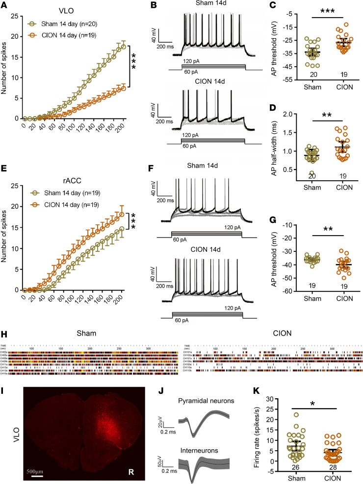
Figure 4. The excitability of VLO excitatory pyramidal neurons decreased in TN mice.
(A) Number of spikes induced by injected currents in VLO CaMK2A+ pyramidal neurons from sham and CION mice. ***P < 0.001, 2-way ANOVA followed by post hoc Student-Newman-Keuls test; n = 20 (sham) and 19 (CION; cells). (B) Examples of AP responses to positive current steps recording from CaMK2A+ pyramidal neurons in the VLO from sham and CION mice. (C and D) Quantification of AP thresholds (C) and half-width (D) in VLO CaMK2A+ pyramidal neurons from sham and CION mice. **P < 0.01, ***P < 0.001, 2-sided Student’s t test; n = 20 (sham) and 19 (CION; cells). (E) Number of spikes induced by injected currents in rACC CaMK2A+ pyramidal neurons from sham and CION mice. ***P < 0.001, 2-way ANOVA followed by post hoc Student-Newman-Keuls test; n = 19 (both sham and CION; cells). (F) Examples of AP responses to positive current steps recording from CaMK2A+ pyramidal neurons in the rACC from sham and CION mice. (G) Quantification of AP thresholds in rACC CaMK2A+ pyramidal neurons from sham and CION mice. **P < 0.01, 2-sided Student’s t test; n = 19 (both sham and CION; cells). (H) Examples of multiple channel recordings in vivo during TST in 1 sham and 1 CION mice. (I) Photomicrograph of coronal section showing the site of multiple-channel electrode implantation in unilateral VLO (contralateral to the CION). Scale bar: 500 μm. (J) Example showing that VLO pyramidal neurons has a long duration compared with interneurons in multiple channel electrophysiological recordings in vivo. (K) The spontaneous firing rate of VLO pyramidal neurons in TN mice was lower during the TST. *P < 0.05, 2-sided Student’s t test; n = 26 (sham) and 28 (CION; cells).

