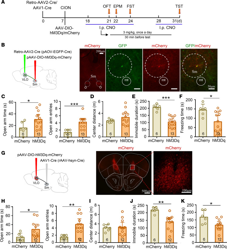
Figure 9. Chemogenetic activation of Sm-VLO projection pathway resulted in the antianxiodepressive effect in TN mice.
(A) Schematic of the protocol for experiments in B–K. (B) Sagittal schematic diagrams showing retro-AAV2-CRE-GFP injection into the bilateral VLO and AAV-DIO-hM3Dq-mCherry (or AAV-DIO-mCherry) injection into the bilateral Sm in mice. Photomicrograph of coronal section showing Cre-dependent mCherry+ signals in the bilateral Sm (low magnification) and both retrograde labeled and Cre-dependent mCherry double-labeled neurons in the Sm (higher magnification). Scale bar: 200 μm for low magnification, 50 μm for high-magnification. (C–F) Activation of the Sm-VLO projection pathway by chemogenetic manipulation produced an antianxiodepressive effect in EPM (C), FST (E), and TST (F), but not in OFT (D). *P < 0.05, ***P < 0.001, 2-sided Student’s t test; n = 6 (mCherry) and 13 (hM3Dq). (G) Sagittal schematic diagrams and photomicrograph of coronal section showing AAV1-Cre injection into the bilateral Sm and AAV-DIO-hM3Dq-mCherry (or AAV-DIO-mCherry) injection of the bilateral VLO in mice. Scale bar: 500 μm for low magnification, 100 μm for high magnification. (H–K) Chemogenetic activation of the VLO neurons receiving projection from Sm produced an antianxiodepressive effect in EPM (H), FST (J), and TST (K), but not in OFT (I). *P < 0.05, **P < 0.01, 2-sided Student’s t test; n = 6 (mCherry) and 12 (hM3Dq).

