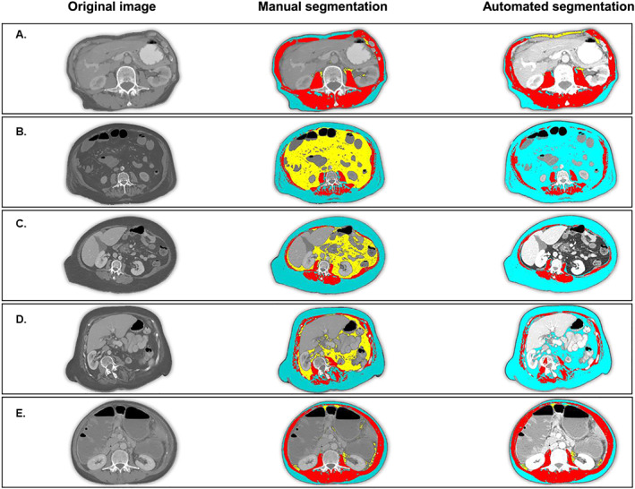
Figure 4.

Cases of segmentation failure: Emaciated body habitus and anatomic abnormalities can cause segmentation failure because ABACS relies on the characteristic shape of the L3 muscle groups. Blue = subcutaneous adipose tissue; red = skeletal muscle tissue; yellow = visceral adipose tissue. In each case of segmentation failure, the abdominal muscle wall is atypical (not continuous or asymmetrical) and thus cannot be identified by the shape‐based prior upon which the ABACS algorithm relies. (A) Patient with emaciated body habitus, in which the thin subcutaneous adipose tissue layer and proximity of the organs to the abdominal muscle wall cause ABACS to mislabel visceral adipose tissue. Jaccard scores for muscle and visceral and subcutaneous adipose tissue are 75, 17, and 73, respectively. (B) Patient in whom muscle wasting (including substantial inter‐muscular adipose tissue) interrupt the continuity of the anterior abdominal wall, causing ABACS to fail to delineate subcutaneous from visceral adipose tissue. Jaccard scores for muscle and visceral and subcutaneous adipose tissue are 80, 0, and 39, respectively. (C) Proximity of organs and bulge in abdominal muscle wall cause visceral adipose tissue segmentation failure. Jaccard scores for muscle and visceral and subcutaneous adipose tissue are 76, 0, and 93, respectively. (D) Patient with scoliosis. Shape of abdominal muscles is not symmetrical and cannot be identified by shape‐based prior segmentation. Jaccard scores for muscle and visceral and subcutaneous adipose tissue are 63, 0, and 69, respectively. (E) Image lacks a continuous musculature in the anterior abdominal wall leading to misclassification of visceral adipose tissue. Jaccard scores for muscle and visceral and subcutaneous adipose tissue are 81, 54, and 81, respectively.
