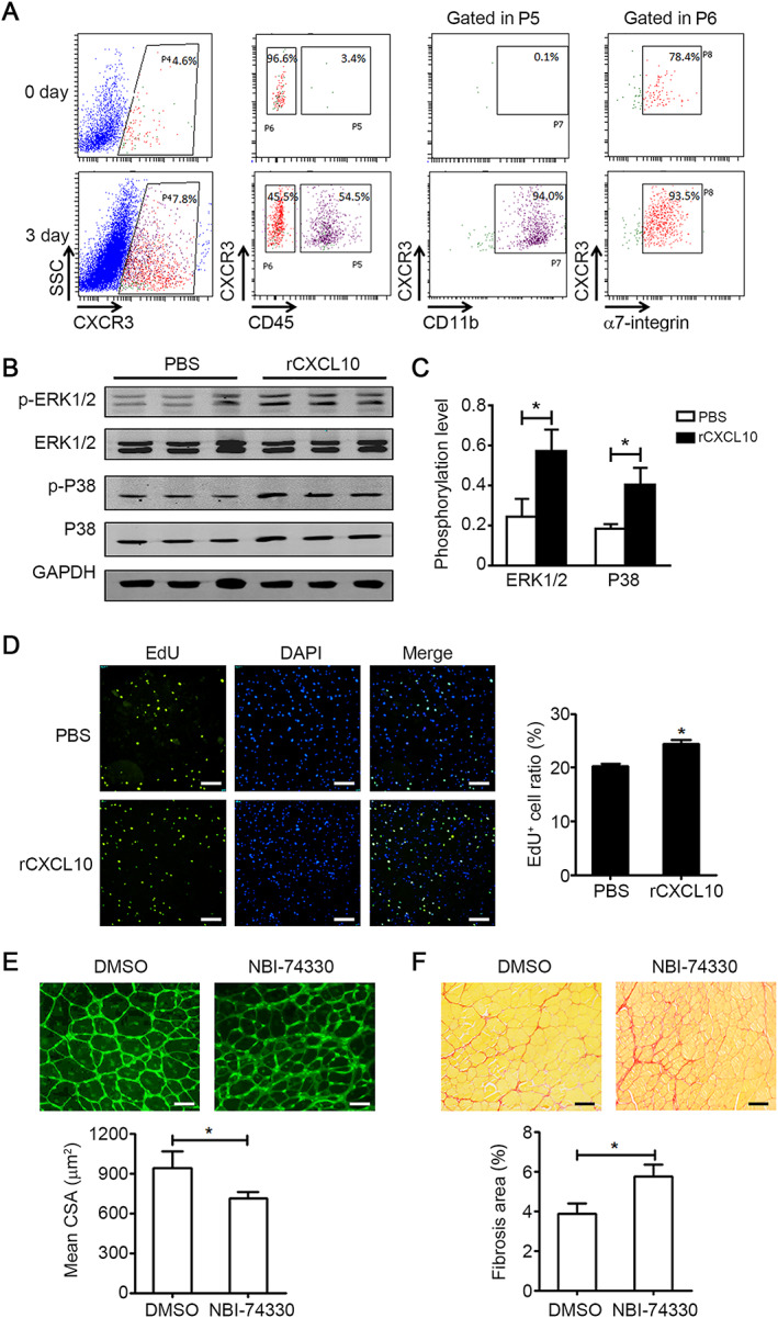
FIGURE 5.

CXCL10 promotes the proliferation and differentiation of muscle satellite cells (MuSCs). (A) Flow cytometry analysis of CXCR3+ cells in young muscle at day 0 and day 3 after injury. (B and C) Primary myoblasts were treated with either phosphate‐buffered saline (PBS) or rCXCL10 (20 ng/mL) for 30 min; and phospho‐ERK1/2 (p‐ERK1/2), phospho‐MAPKp38 (p‐P38), total ERK1/2, and total MAPKp38 (P38) protein levels were quantified by western blot (B) and normalized to GAPDH (C) (n = 3 per group). (D) Quantification of the ratio of EdU+ primary myoblasts cultured for 24 h in medium with either PBS or rCXCL10 (20 ng/mL) (bars = 100 μm, n = 6 per group). (E) CXCR3 antagonist NBI‐74330 (100 mg/kg) or DMSO was administered i.v. at 1 and 3 days after injury. The mean myofibre cross‐section area (CSA) was accessed by wheat germ agglutinin (WGA) staining at 15 days after injury (bars = 50 μm, 150~200 myofibre per muscle were examined, n = 4 per group). (F) At day 15 after injury, the ratio of Sirus Red positive fibrosis area in muscle from muscle with NBI‐74330 or DMSO treatment (bars = 100 μm, n = 4 per group) was quantified. Data represent the mean ± SEM. *P < 0.05, by two‐tailed Student's t‐test.
