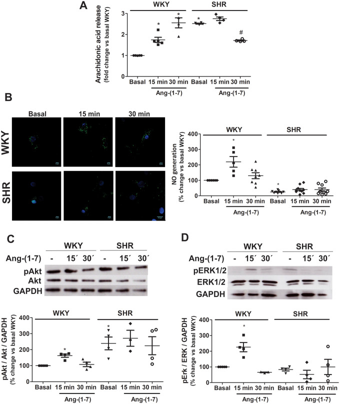
Figure 2.
Ang-(1-7) responses are blunted in SHR neurons. AA release (A), NO generation (B), Akt phosphorylation (C), and ERK1/2 phosphorylation (D) were measured in brainstem neurons from WKY rats and SHRs incubated in the absence (basal) or presence of Ang-(1-7). Representative images of NO (NO in green and nucleus in blue) generation and representative western blots of total and phosphorylated Akt and ERK1/2 are presented in panels (B–D), respectively. The results are presented as changes in response relative to WKY neurons in the basal condition. The line in each scatter plot represents the mean ± SEM of four independent neuronal cultures. Each experiment was carried out with 3.5 million (A), 500 000 (B) and eight million cells (C and D). *P < 0.05 vs. basal WKY; #P < 0.05 vs. basal SHR (Kruskal–Wallis non-parametric tests followed by Dunn’s multiple comparison tests).

