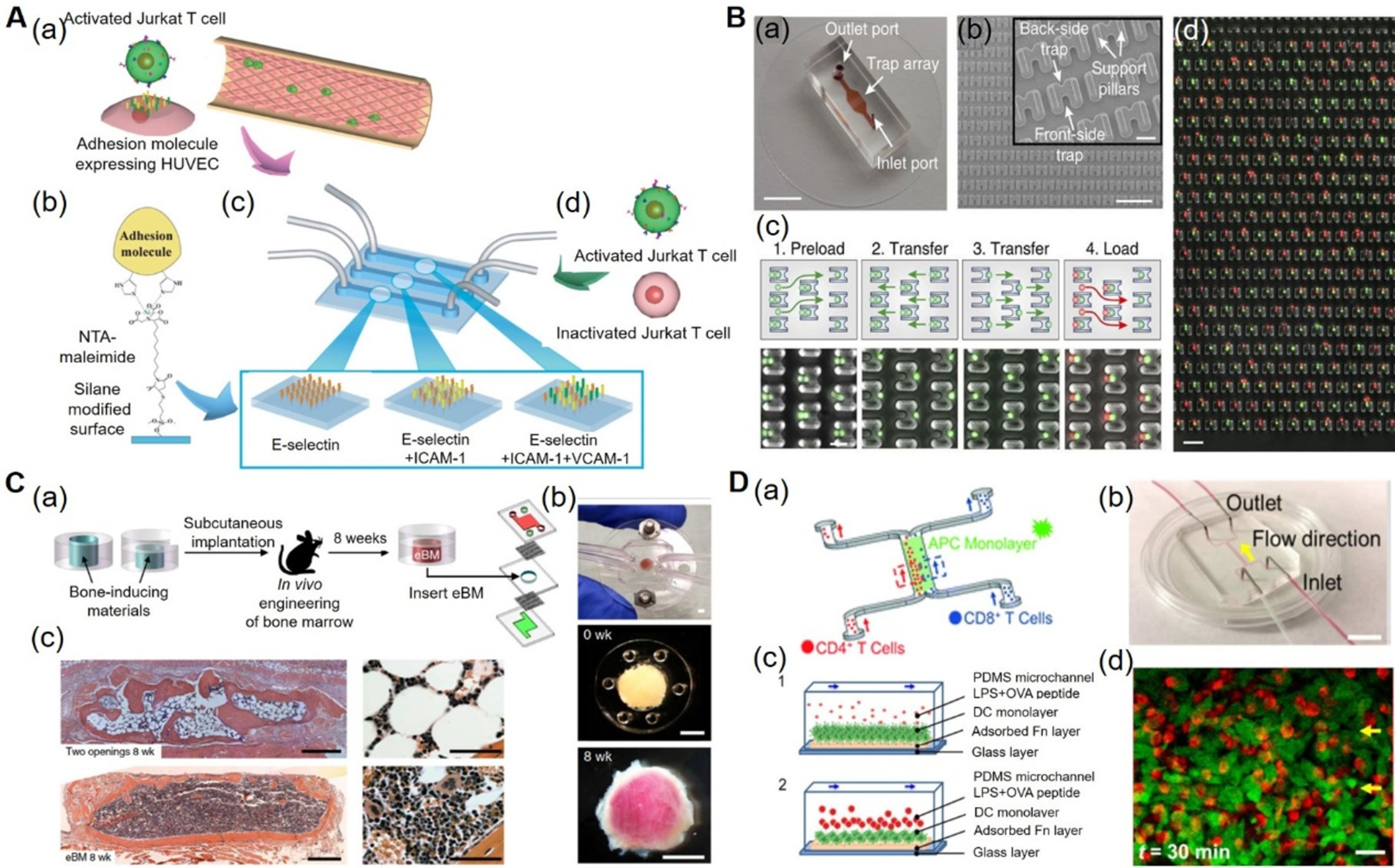
Figure 2. Immune OOC models.

A) Inflammatory-mimetic microfluidic chip. (a) Schematic of the Inflammatory mimetic microfluidic chip. Activated Jurkat T-cells and HUVECs. (b) Adhesion molecules such as E-selectin, ICAM-1, and VCAM-1 were immobilized in the fluidic channel by treating with MPTMS and NTA-maleimide. (c) Schematics of vascular endothelia formed with adhesion molecule-expressing HUVECs. (d) Infusion of activated or drug-treated Jurkat T-cells into the channels to study the T-cell interactions. Reproduced with permission.[45] Copyright 2014, Royal Society of Chemistry. B) Microfluidic device for immune cell pairing. (a) Photograph of microfluidic cell-pairing device. (b) Scanning electron micrographs of the cell trap. (c) Cell-loading and pairing protocol. (d) Phase-contrast and fluorescence images showing pairing of primary mouse lymphocytes (red) and DCs (green) in the traps. Reproduced under the Creative Commons Attribution 4.0.[49] International Public License for open access articles. C) Bone marrow-on-a-chip. (a) Schematics of a bone marrow-on-a-chip in which eBM within a PDMS device formed in vivo was later explanted and cultured in a microfluidic system. (b) Bone marrow-on-a-chip microdevice used to culture the eBM in vitro (top), the PDMS device containing the matrix and bone-inducing factors before implantation (middle), and the white cylindrical bone with pink marrow visible within the eBM explanted after 8 weeks (bottom). (c) Hematoxylin and eosin staining of the eBM formed in the PDMS device with two openings (top) and one opening (bottom) at 8 weeks of implantation. Reproduced with permission.[63] Copyright 2014, Springer Nature. D) Immune cell-cell interaction-on-a-chip. (a) Schematics of the immune cell-cell interaction-on-chip. (b) The PDMS chip consisting of one main flow channel for cells with two inlets and two outlets. (c) Schematics showing the dynamic interactions between DCs and T-cells, in the presence of LPS and OVA peptides (1) and loading of T-cells and their interactions with DCs (2). (d) Confocal images showing the T-cell and DC interactions within 10 mins and 30 mins. Reproduced with permission.[78] Copyright 2016, Royal Society of Chemistry. DCs: dendritic cells, eBM: engineered bone marrow, HUVECs: human umbilical vein endothelial cells, ICAM-1: intercellular adhesion molecule-1, LPS: lipopolysaccharides, MPTMS: (3-mercaptopropyl)trimethoxysilane, NTA-maleimide: nitrilotriacetic acid-maleimide, OVA: ovalbumin, PDMS: polydimethylsiloxane, OOC: organ-on-a-chip, VCAM-1: vascular cell adhesion molecule-1.
