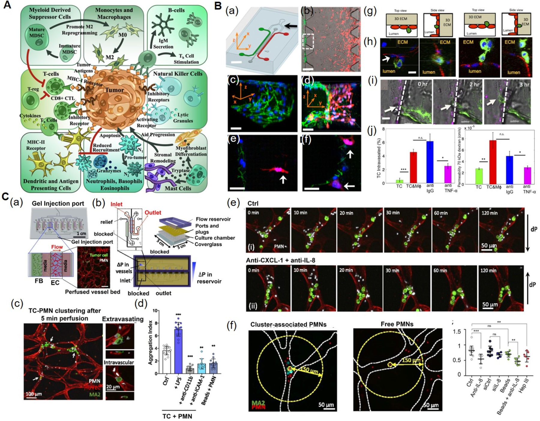
Figure 4. Immune microenvironments in tumors and in vitro tumor-on-a-chip models.

A) The TME comprises of tumor cells, as well as cellular components of both the innate and adaptive immune systems. Reproduced under the Creative Commons Attribution License 4.0.[132] International License for open access articles. B) Microfluidic tumor-vascular interface model. (a) Endothelial channel (green), tumor channel (red), and 3D ECM (dark gray). b) Phase-contrast image showing the invasion of fibrosarcoma cells (HT1080, red) through the ECM (gray) towards the endothelium (MVECs, green). (c) VE-cadherin and nuclei staining showing the endothelial monolayer. (d) 3D rendering of a confocal image showing the invasion of tumor cells and adhesion to the endothelium. (e) Invasion of HT1080 cells (white arrow) towards the endothelium. (f) HT1080 cells attached to the endothelial monolayer. (g) Schematics showing the endothelial layer, the tumor cells, and the 3D ECM within the device. (h) Confocal images showing intravasation of a MDA-MB-231 cell (green) across the MVEC layer (stained red for VE-cadherin). (i) Time lapse confocal images showing the intravasaton of a MDA-MB-231 cell (white arrow) across a HUVEC monolayer (magenta) in the presence of RAW264.7. (j) Intravasation of MDA-MB-231 cells and endothelial permeability of 70 kDa dextrans across a HUVEC monolayer in the presence of macrophages. Blocking of macrophage-secreted TNF-α resulted in a significant reduction in both intravasation and permeability compared to the IgG antibody control. Reproduced under the Creative Commons Attribution-NonCommercial-NoDerivatives 4.0.[139] International Public License for open access articles. C) Multiplexed microvasculature device for dynamic inflamed neutrophil and the tumor cell interactions. (a) Schematics of device consisting of eight independent hydrogel regions where microvascular networks are formed. Inset depicts a confocal image of one perfused vascular network. (b) Schematics and photographs showing different components of the device. (c) PMN and TC clusters in microvessels, highlighting extravasating and nonextravasated A375-MA2 cells in TC-PMN clusters. (d) Degree of TC-PMN aggregate formation during intravascular arrest. (e) The dispersion of cluster-associated PMNs from TCs over 2 h, in the absence and presence of anti-CXCL-1+anti-IL-8. (f) Cluster-associated and free PMNs arrested intraluminally, and fraction of PMNs remaining in individual clusters over 6 h in the presence of IL-8 and/or CXCL-1 neutralizing antibodies, or when tumor cells are replaced with 15 μm polystyrene beads. Reproduced under the Creative Commons Attribution-NonCommercial-NoDerivatives 4.0.[141] International Public License for open access articles. CXCL-1: chemokine (C-X-C motif) ligand-1, 3D: three-dimensional, HUVECs: human umbilical vein endothelial cells, IL-8: interleukin-8, IgG: immunoglobulin G, MVECs: human microvascular endothelial cells, PMN: LPS-stimulated neutrophils, TC: tumor cell, TME: tumor microenvironment, TNF-α: tumor necrosis factor-alpha.
