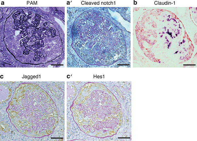
Figure 3 |. Aberrant Notch expression is observed in hyperplastic parietal epithelial cells (PECs) in human collapsing focal segmental glomerulosclerosis.
(a) Periodic acid-silver methenamine (PAM) staining showed severely collapsed glomerular capillaries with extracapillary hyperplasia. On serial sections, cleaved Notch1 staining (a’, brown dots) was observed in hyperplastic epithelial cells but was almost completely absent in glomerular tufts. (b) Extracapillary hyperplastic epithelial cells expressed claudin-1 (red) but were negative for Nestin (violet). On serial sections, Jagged1 (c) and Hes1 (c’) colocalized in hyperplastic PECs (brown), as shown with periodic acid–Schiff (PAS) counterstaining. Bars = 40 μm.

