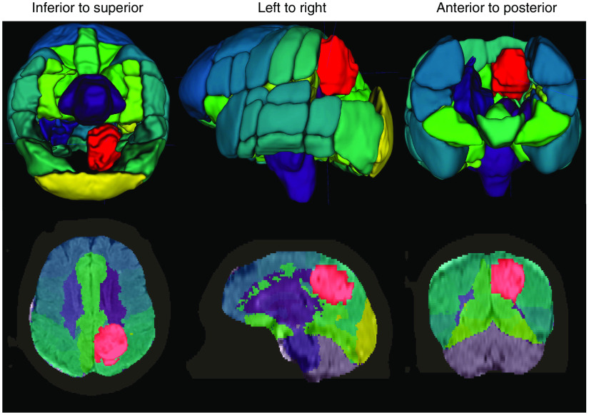
Fig. 6.
Example output from our DL system for automatic brain tumor segmentation. The system loads an MRI exam containing a T1-weighted postcontrast scan (T1c) and a FLAIR scan. Input from a wide range of MRI scanners and with varying scan parameters will work. We designed the system to perform the following steps automatically, without additional input: (1) enhance the MRI scans to remove artifacts; (2) identify the brain within the MRI scan (strip the skull), even in the presence of significant pathology or surgical interventions; (3) segment the brain tumor; and (4) coregister the Harvard-Oxford probabilistic atlas to the brain. The last step is used for visualization purposes and is optional. In this image, the tumor is red. Other colors indicate various atlas regions. The top and bottom rows show 3D and 2D views of the output data, respectively. Several atlas regions in the vicinity of the tumor have been made transparent in the 3D view to aid tumor visualization.

