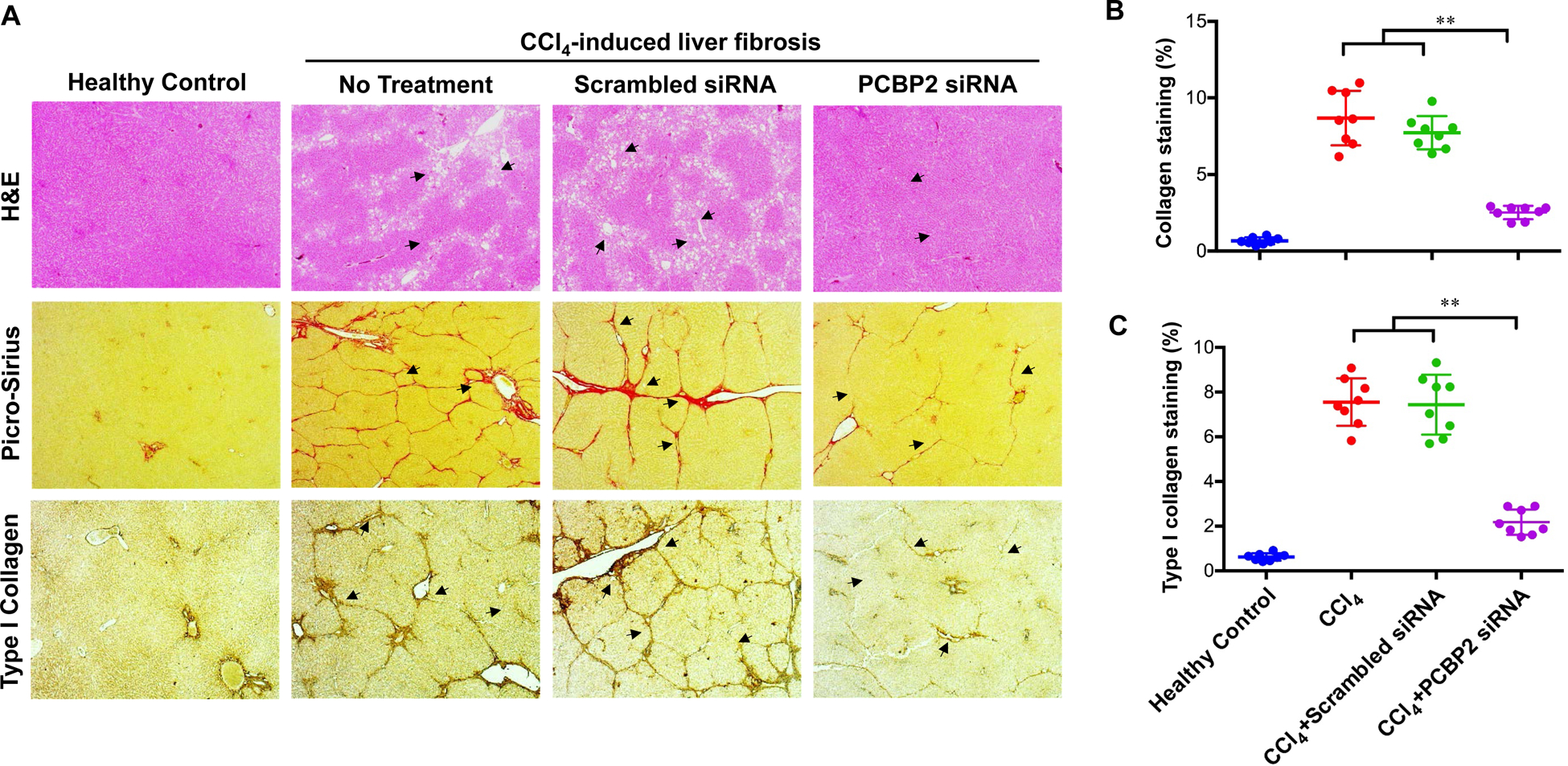
Figure 7. Histological analysis of liver specimen.

(A) H&E, Picro-Sirius Red, and type I collagen immunohistochemistry staining of liver specimen; (B) Picro-Sirius Red stained areas were quantified with ImageJ; (C) Type I collagen positive areas were quantified with ImageJ. (n=8) (** p<0.01)
