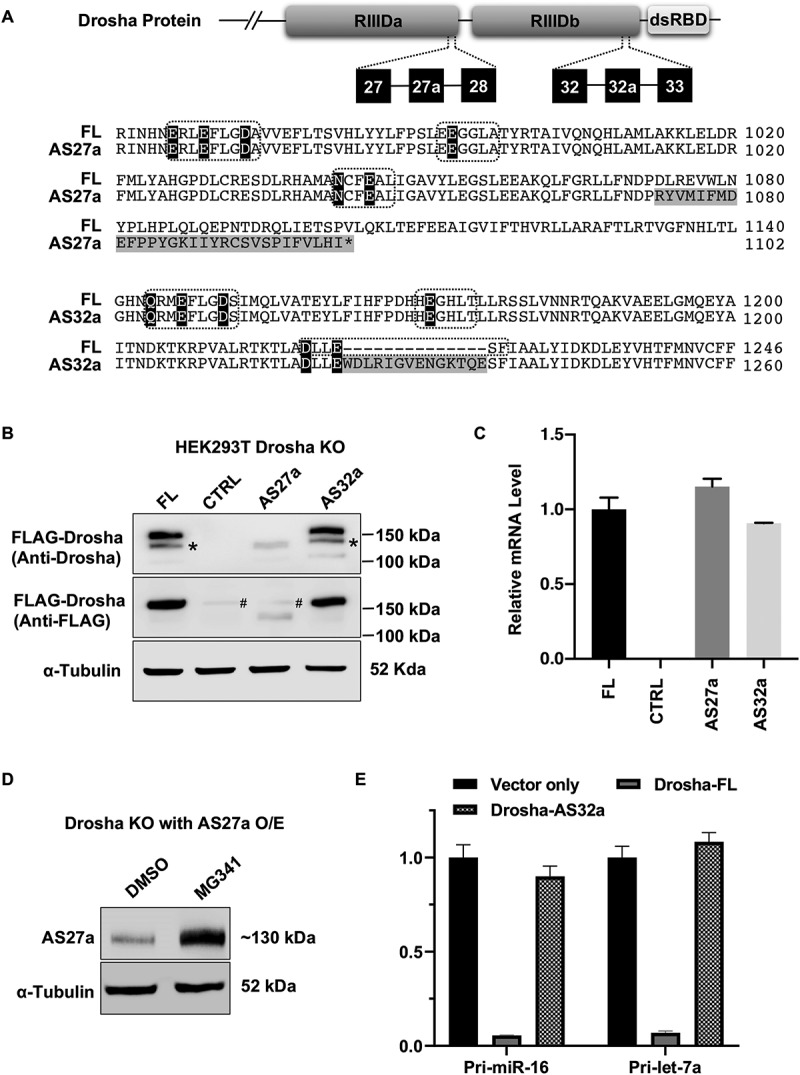
Figure 2.

Characterization of Drosha-AS27a and Drosha-AS32a isoforms.
(A) Sequence alignment of amino acids encoded by Drosha-FL and alternative splice variants. Upper panel indicates where exon 27a and exon 32a are located in the Drosha protein domain. In the lower panel, sequence differences between amino acids in Drosha-FL and Drosha-AS27a, or between Drosha-FL and Drosha AS32 are highlighted in grey. The signature motifs of the RNase III domain are indicated by the dashed-line box. Amino acid residues critical for cleavage activity are highlighted in black. (B) Western blot detecting protein expression of Drosha alternative splicing variants in Drosha KO cells. As previously described, N-terminal truncated Drosha resulting from degradation was also detected and labelled with *. Unspecific bands for anti-FLAG antibody were labelled with #. (C) RT-qPCR was used to quantitate mRNA levels of Drosha splicing variants. (D) Western Blot detecting ectopic expression of Drosha-AS27a in Drosha KO cells upon treatment with either DMSO or Bortezomib (MG341, 200 nM) for 12 hours. (E) Dual-luciferase assays to measure Drosha cleavage activity. Cleavage reporters containing either pri-miR-16-1 or pri-let-7a-1 were co-transfected with empty vectors or plasmids expressing Drosha-FL or Drosha-AS32a into Drosha KO cells. See more details in Supplementary Fig. S4.
