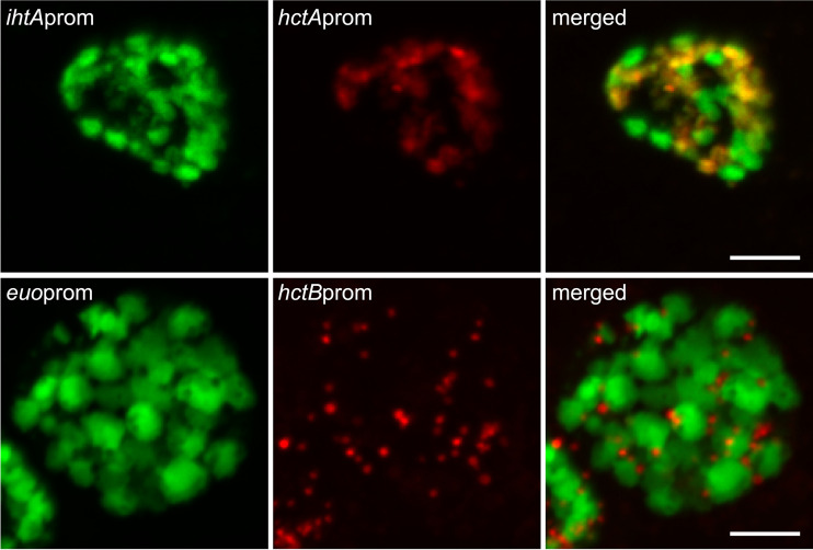
FIG 9.
Confocal fluorescence microscopy of cell type promoter expression upon inhibition of chlamydial division. Host cells were infected with Ctr-hctAprom-mKate2/ihtAprom-mNeonGreen (red and green, respectively) or Ctr-hctBprom-mKate2/euoprom-Clover (red and green, respectively), followed by treatment with penicillin (Pen) at 20 hpi. Samples were fixed at 24 hpi. Fixed samples were imaged by confocal microscopy, and maximum intensity projections are shown. Scale bars, 5 μm.

