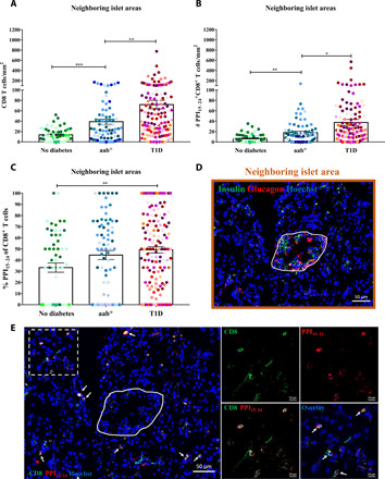
Fig. 2. PPI15–24-specific CD8+ T cells are attracted to the proximity of and into the islets.

Neighboring islet areas (n = 267) were randomly selected from pancreas tissue sections of all 22 donors (no diabetes, n = 57; aab+, n = 73; and T1D, n = 137). (A) A higher density of CD8+ T cells close to the islets in aab donors compared with donors without diabetes (P = 0.0004). (B) The number of PPI15–24+CD8+ T cells is increased in donors with abb+ (P = 0.089) and T1D (P < 0.0001) compared with donors without diabetes. (C) Higher frequency of PPI15–24-specific CD8+ T cells in neighboring islet areas in donors with T1D compared with donors without diabetes (P = 0.0061). Every dot represents a neighboring islet area. Bars represent the mean ± SEM values in different groups. Every color represents a donor. PPI15–24-specific CD8+ T cells were counted manually, and the density was calculated per square millimeter. For statistical analysis, nonparametric Kruskal-Wallis test followed by Dunn multiple comparison test was used to determine significance: *P = 0.05, **P < 0.05, and ***P < 0.001. (D) Restaining of a pancreas tissue section from a donor with aab+ (#6154) for insulin and glucagon (see Supplementary Methods for more details). The image shows an insulin-containing islet. The neighboring islet area is demonstrated in brown. (E) PPI15–24-specific CD8+ T cells were found close to islets already in donors with aab+. White arrows indicate PPI15–24-specific CD8+ T cells.
