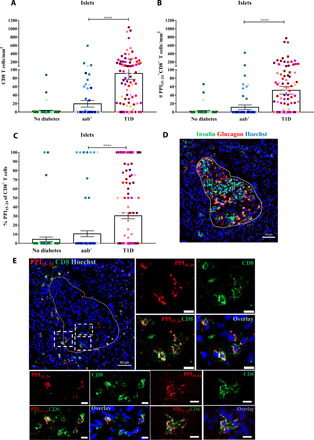
Fig. 3. PPI15–24-specific CD8+ T cells are found within the islets of donors with T1D.

Islets were randomly selected from pancreas sections of donors without diabetes (n = 63), aab+ (n = 83), and T1D (n = 156). (A) A higher density of CD8+ T cells in donors with T1D (P < 0.0001) compared with donors with aab+. (B) High numbers of PPI15–24-specific CD8+ T cells in donors with T1D (P < 0.0001). (C) The percentage of PPI15–24-specific CD8+ T cells in the islets is higher in donors with T1D. Every dot represents an islet (n = 302). Bars represent the mean ± SEM values. For statistical analysis, nonparametric Kruskal-Wallis test followed by Dunn multiple comparison test was used to determine significance: ****P < 0.0001. (D and E) Representative immunofluorescence images of a pancreas section from a donor with T1D (#6052, 1 year of disease duration). (D) Restaining for insulin and glucagon (see Supplementary Methods for more details). The image shows an insulin-containing islet. (E) In situ PPI15–24 staining (red) combined with CD8 (green) and nuclear marker (blue, Hoechst). PPI15–24-specific CD8+ T cells are shown in yellow. Magnification ×20. Scale bars, 10 μm for cropped images.
