. 2020 Oct 16;9:e55542. doi: 10.7554/eLife.55542

Figure 7. Methylation of the viral DNA and its suppression by V2 occur in a Cajal body-dependent manner.

(A) V2-GFP co-localizes with Fibrillarin-RFP (nucleolus and Cajal body marker) in the Cajal body, while GFP-V2 does not. RFP-Fibrillarin and V2-GFP or GFP-V2 were transiently co-expressed in N. benthamiana epidermal cells. Confocal images were taken at two days after infiltration. Arrowheads indicate the position of the Cajal body. Bar, 5 μm. This experiment was repeated three times with similar results. (B) Quantification of Cajal body localization of V2-GFP or GFP-V2. (C) V2-GFP, but not GFP-V2, can restore the suppression of viral DNA methylation of a V2 null TYLCV mutant in local infection assays. Cytosine methylation in the intergenic region (IR) of the V2 null mutant genome in locally infected leaf patches of N. benthamiana expressing V2, V2-GFP, or GFP-V2 was detected by bisulfite sequencing at 3 days post-inoculation (dpi). TYLCV or TYLCV-V2null alone were used as controls. The original single-base resolution bisulfite sequencing data are shown in Figure 7—figure supplement 1B. This experiment was repeated twice with similar results. (D) Nbcoilin expression in Nbcoilin-silenced (TRV-Nbcoilin) and control plants (TRV-EV) infected with TYLCV, TYLCV-V2null, or mock-inoculated at 3 wpi measured by RT-qPCR. Gene expression was normalized to NbTubulin. Values are the mean of six independent biological replicates; error bars indicate SEM. Asterisks indicate a statistically significant difference according to Student’s t-test. **: p<0.01. (E) Percentage of methylated cytosines in the intergenic region (IR) of TYLCV in systemic infection assays with TYLCV wild-type or V2 null mutant (TYLCV-V2null) in Nbcoilin-silenced (TRV-Nbcoilin) or control (TRV-EV) N. benthamiana plants at 3 weeks post-inoculation (wpi), as detected by bisulfite sequencing. Values are the mean of three independent biological replicates; error bars indicate SEM. The original single-base resolution bisulfite sequencing data are shown in Figure 7—figure supplement 2D.

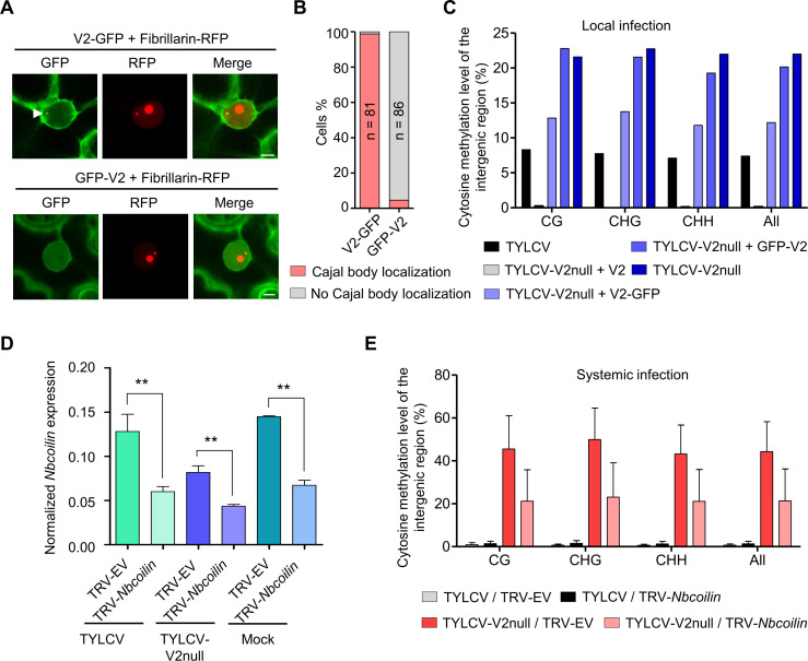
Figure 7.

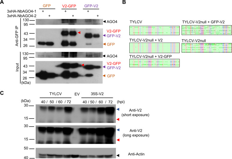
Figure 7—figure supplement 1. Relevance of the Cajal body localization of the V2-AGO4 interaction.

Figure 7—figure supplement 1.

(A) 3xHA-NbAGO4-1 and 3xHA-NbAGO4-2 interact with V2-GFP and GFP-V2 in co-immunoprecipitation (co-IP) assays upon transient expression in N. benthamiana. Free GFP was used as negative control. Three independent biological replicates were performed with similar results. (B) Original single-base resolution bisulfite sequencing data for Figure 7C. >16 individual clones were sequenced per sample and replicate. Each single circle, corresponding to a cytosine, is colored in blue, red, or green, representing the CHG, CG or CHH contexts, respectively. Methylated cytosines are represented by filled circles, while unmethylated cytosines are represented by empty circles. (C) V2 protein levels in N. benthamiana leaves locally infected by TYLCV or transiently expressing V2 under the 35S promoter. Empty vector (EV) was used as negative control. Three independent biological replicates were performed with similar results. Red arrowheads indicate the predicted size of the V2 protein; blue arrowheads indicate an additional specific band of higher size. hpi, hours post-inoculation.
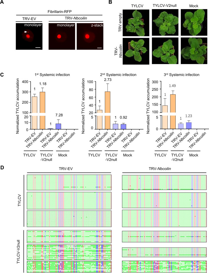
Figure 7—figure supplement 2. TYLCV infection in Nbcoilin-silenced plants.

Figure 7—figure supplement 2.

(A) No Cajal body was observed in the nuclei of Nbcoilin-silenced plants, whereas in control plants at least one Cajal body was normally present in each nucleus; these results are in agreement with Shaw et al., 2014. Fibrillarin-RFP, used as a nucleolus and Cajal body marker, was transiently expressed in N. benthamiana epidermal cells of Nbcoilin-silenced (TRV-Nbcoilin) and control plants (TRV-EV). Confocal images were taken at 2 days after infiltration. Arrowheads indicate the position of the Cajal body. Bar, 5 μm. This experiment was repeated three times with similar results. (B) Representative pictures of N. benthamiana plants infected with the indicated combinations of viruses. Photographs were taken at 3 weeks post-inoculation (wpi). (C) Viral (TYLCV) accumulation in systemic infections in Nbcoilin-silenced or control plants, measured by qPCR. Apical leaves from six plants were collected at 3 wpi. The experimental design is shown in Figure 3—figure supplement 1B. The accumulation of viral DNA is normalized to the 25S ribosomal RNA interspacer (ITS). Results from three independent experiments are shown. Values are the mean of six independent biological replicates; error bars indicate SEM. The relative fold change of viral accumulation between Nbcoilin-silenced plants and control plants is shown above each column. (D) Original single-base resolution bisulfite sequencing data for Figure 7E. >11 individual clones were sequenced per sample and replicate. Each single circle, corresponding to a cytosine, is colored in blue, red, or green, representing the CHG, CG, or CHH contexts, respectively. Methylated cytosines are represented by filled circles, while unmethylated cytosines are represented by empty circles. Values of IR methylation in independent biological replicates are shown in Supplementary file 1.
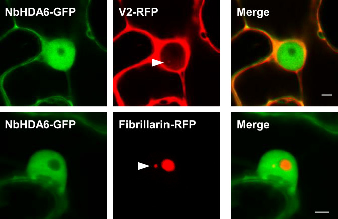
Figure 7—figure supplement 3. NbHDA6-GFP does not localize to the Cajal body.

Figure 7—figure supplement 3.

NbHDA6-GFP and V2-RFP or Fibrillarin-RFP were transiently co-expressed in N. benthamiana epidermal cells. Fibrillarin-RFP is used as nucleolus and Cajal body marker. Confocal images were taken at 2 days after infiltration. Arrowheads indicate the position of the Cajal body. Bar, 5 μm. This experiment was repeated three times with similar results.
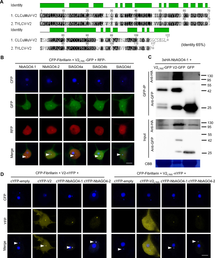
Figure 7—figure supplement 4. TYLCV V2L76S interacts with AGO4 in the Cajal body.

Figure 7—figure supplement 4.

(A) Alignment of the amino acid sequences of V2 from Cotton Leaf Curl Multan virus (CLCuMuV) and V2 from TYLCV. The alignment was performed by Geneious (https://www.geneious.com). Black background indicates conservation. The identity of these two proteins is 65%. (B) V2L76S-GFP and RFP-AGO4 co-localize in the Cajal body. CFP-Fibrillarin, V2L76S-GFP, and RFP-NbAGO4-1/2 or RFP-SlAGO4a/b/d were transiently co-expressed in N. benthamiana epidermal cells. CFP-Fibrillarin is used as anucleolus and Cajal body marker. Confocal images were taken at two days after infiltration. Arrowheads indicate the position of the Cajal body. Bar, 5 μm. This experiment was repeated three times with similar results. (C) 3xHA-NbAGO4-1 interacts with V2-GFP and V2L76S-GFP in co-immunoprecipitation (co-IP) assays upon transient expression in N. benthamiana. Free GFP was used as negative control. CBB, Coomassie brilliant blue staining. The V2-GFP sample was diluted 1/20 for western blot to reach a protein amount comparable to that of V2L76S-GFP. Three independent biological replicates were performed with similar results. (D) V2L76S interacts with AGO4 in the Cajal body. The N-terminal half of the YFP fused to V2L76S (V2L76S-nYFP) was transiently co-expressed with the C-terminal half of the YFP alone (cYFP, as negative control), or cYFP-NbAGO4, cYFP-SlAGO4, or cYFP- V2L76S in N. benthamiana leaves. CFP-Fibrillarin was used as a nucleolus and Cajal body marker. V2 was used as control. Yellow fluorescence indicates a positive interaction. Arrowheads indicate the position of the Cajal body. Bar, 5 μm. This experiment was repeated three times with similar results.
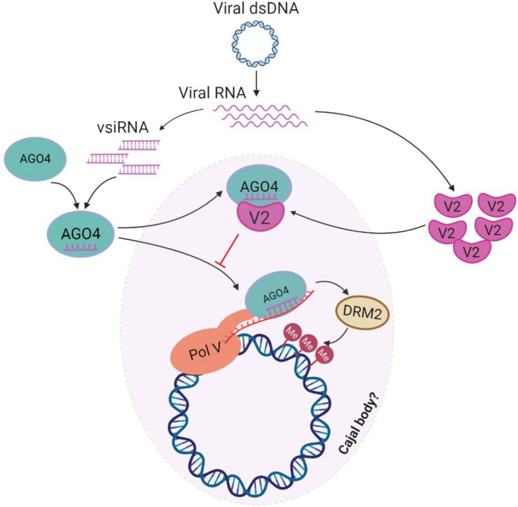
Figure 7—figure supplement 5. Model for the V2-mediated inhibition of the AGO4-dependent methylation of the viral DNA.

Figure 7—figure supplement 5.

During the viral infection, the ssDNA TYLCV genome forms dsDNA replicative intermediates, which could be targeted by the host AGO4-dependent RNA-directed DNA methylation (RdDM) pathway in a Cajal body-dependent manner as an antiviral defence mechanism. Viral small interfering RNA (vsiRNA) are generated and loaded into AGO4. In the absence of the virus-encoded V2 protein, the AGO4-vsiRNA complex could be effectively guided toward the viral genome by complementary base pairing to the scaffold RNA and association with Pol V, and recruit the methyltransferase DRM2 to catalyze methylation of the viral genome. When V2 is present, however, V2 interacts with AGO4 and interferes with the binding of this protein to the viral RNA and the viral DNA, enabling viral evasion from the AGO4-dependent DNA methylation.
Figure 7—video 1. Z-stack video showing the co-localization of V2-GFP and Fibrillarin-RFP in the Cajal body.
Download video file (5.6MB, mp4)
Figure 7—video 2. Z-stack video showing the localization of GFP-V2 and Fibrillarin-RFP.
Download video file (6.2MB, mp4)