. 2020 Sep 21;9:e55589. doi: 10.7554/eLife.55589

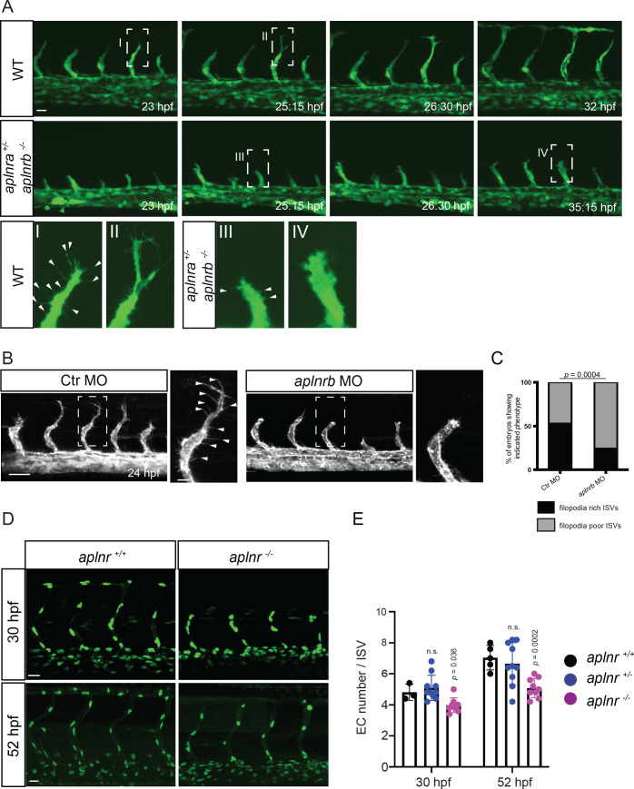
Figure 2. Apelin signaling regulates endothelial filopodia formation and endothelial cell numbers.

(A) Still images from confocal time-lapse movies of vascular development in wild-type and aplnra +/-; aplnrb -/- embryos. During sprouting, wild-type tip cells send out filopodia (arrowheads). aplnra +/-; aplnrb -/- embryos exhibit smaller sprouts and fail to form filopodia. (B) Confocal images of the blood vasculature in 24 hpf Tg(kdrl:HsHRAS-EGFP) embryos injected with Ctr MO and aplnrb MO. aplnrb morphant embryos exhibit smaller sprouts and fail to form filopodia (arrowheads). (C) aplnrb morphant embryos exhibit a reduction in the number of endothelial filopodia (Ctr MO, n = 10; aplnrb MO, n = 15). (D) Confocal images of the blood vasculature of 30 and 52 hpf Tg(fli1a:nEGFP) wild-type and aplnra +/-; aplnrb -/- embryos showing EC cell nuclei. (E) aplnra +/-; aplnrb -/- embryos exhibit reduced EC numbers in the ISVs (30 hpf: aplnr +/+, n = 3; aplnr +/-, n = 10; aplnr -/-, n = 8; 52 hpf: aplnr +/+, n = 5; aplnr +/-, n = 10; aplnr -/-, n = 9). n.s. not significant (two-tailed t-test). Scale bars: A, D, 20 µm; B, 40 µm; B, inset 10 µm.

Figure 2.

Figure 2—figure supplement 1. No obvious defects during initiation of EC sprouting in Apelin signaling-deficient embryos.

Figure 2—figure supplement 1.

Confocal projection images of the trunk region of Tg(fli1a:EGFP) embryos at 21 hpf. Sprouting of ECs from the DA does not appear to be altered in Apelin signaling-deficient embryos. Scale bar: 50 µm.
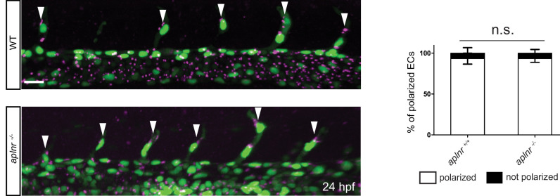
Figure 2—figure supplement 2. No obvious defects in EC polarity in Apelin deficient embryos.

Figure 2—figure supplement 2.

Confocal projection images of the trunk region of Tg(kdrl:NLS-EGFP); Tg(fli1a:B4GALT1-mCherry) embryos at 24 hpf. Apelin deficiency does not cause obvious defects in EC polarity (aplnr +/+, n = 6; aplnr -/-, n = 8). Arrowheads point to polarized tip cells in the ISVs. Scale bar: 20 µm.
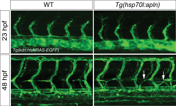
Figure 2—figure supplement 3. Overexpression of apln does not cause ectopic EC sprouting.

Figure 2—figure supplement 3.

Confocal projection images of the trunk region of Tg(fli1a:LIFEACT-GFP) embryos at 23 and 48 hpf. apln expression was induced by a single heatshock at 19.5 hpf for 1 hr at 39°C and analyzed at 23 hpf or heatshocked at 42 and 45 hpf for 1 hr at 39°C and analyzed at 48 hpf. Global overexpression of apln does not lead to ectopic vascular sprouting but mispatterning of the string of parachordal lymphangioblasts (arrows).
Figure 2—video 1. Confocal time-lapse-imaging of a Tg(fli1a:EGFP) wild-type embryo from 23 to 32 hpf.
Download video file (1.4MB, mp4)
During sprouting, wild-type tip cells send out filopodia. Related to Figure 2A.
Figure 2—video 2. Confocal time-lapse-imaging of a Tg(fli1a:EGFP) aplnra +/-; aplnrb -/- embryos from 23 to 35 hpf.
Download video file (1.3MB, mp4)
During sprouting, aplnra +/-; aplnrb -/- embryos exhibit smaller sprouts and fail to form filopodia. Related to Figure 2A.