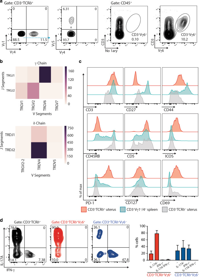
Fig. 2. Surface phenotype, TCR usage, and functional properties of uterine γδ T cells.
a Left: TCR usage of uterine γδ T cells was determined by flow cytometric analysis of Vγ1, Vγ4, and Vγ5 chains. Right: TCR usage of uterine γδ T cells determined by flow cytometry using the Vγ6-specific 1C10-1F7 antibody. Representative flow plots from three experiments are shown. b TCR deep-sequencing analysis of RNA from sorted uterine Vγ1−4−5− cells (representative data from three biological replicates), showing the relative abundance of recombination events for gamma (top) and delta (bottom) gene segments. c The surface immunophenotype of uterine TCRγδ+ and CD3+TCRδ− (i.e. TCRαβ+) T cells, and splenic Vγ1/4+ γδ T cells were determined by flow cytometry. Representative data from two experiments are shown. d Left: Uterine γδ T-cell suspensions were prepared and stimulated with PMA and ionomycin in the presence of Brefeldin A, with IL-17A and IFN-γ production assessed by intracellular staining and flow cytometric analysis in total (left), Vγ6+ (middle, red), and Vγ6− (right, blue) γδ T cells. Right: Percentages of cytokine-secreting cells amongst Vγ6+ (red) and Vγ6− (blue) cells were determined (n = 5 mice). Representative data from two experiments are shown. Graph indicates mean ± SD.

