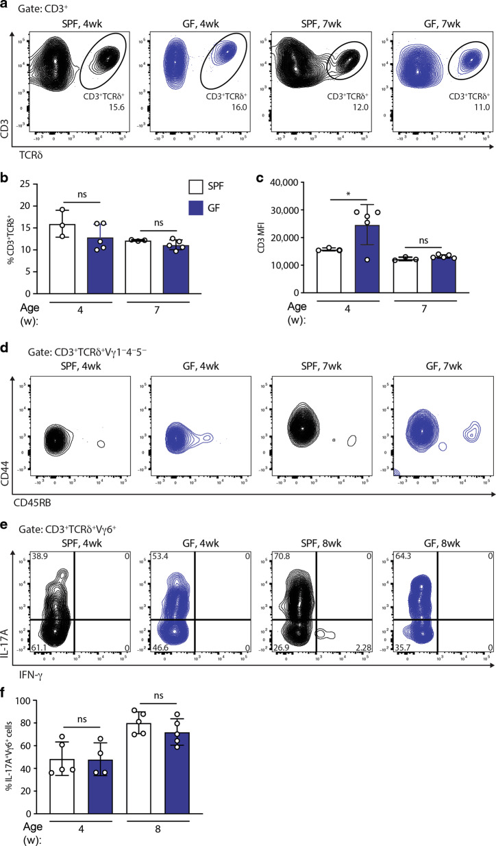
Fig. 4. The microbiome is dispensable for uterine γδ T cells.
a Uterine γδ T-cell staining (gated on CD3+ lymphocytes); b quantification; c CD3 expression; and d CD44 and CD45RB expression in SPF and germ-free (GF) C57BL/6J mice at 4 and 7 weeks (n = 3–5). Representative of four experiments. e Uterine γδ T-cell suspensions from SPF and GF mice were prepared and stimulated with PMA and ionomycin in the presence of Brefeldin A, with IL-17A and IFN-γ production assessed by intracellular staining and flow cytometric analysis in Vγ6+ γδ T cells. f Percentages of IL-17A-secreting cells amongst Vγ6+ cells were determined (n = 4–5 mice). Graph indicates mean ± SD. Statistical significance was assessed by one-way ANOVA with Sidak’s multiple comparisons post-hoc test. ns not significant, *p < 0.05.

