Fig. 5.
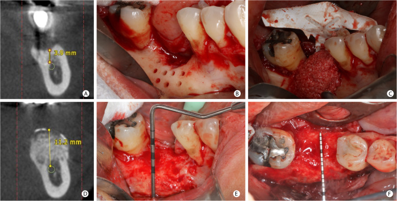
GBR procedure for vertical bone increase in the posterior mandibular region. a Tomographic measurement of the vertical bone defect in the position of the mandibular first molar. b Clinical aspect of a vertical defect after the cortical bone’s perforations with a spiral drill. c Mixture of autogenous and ABBM grafts agglutinated with i-PRF positioned under the d-PTFE-Ti membrane already fixed in the lingual face. d The bone height of 11.2 mm measured on CBCT, after 8.5 months. e Clinical aspect of the vertical bone augmentation. f Occlusal view after GBR

