
Fig. 5. Treatment with 2-DG inhibits glycolysis and stem cell-like characteristics in gastric cancers (GCs).
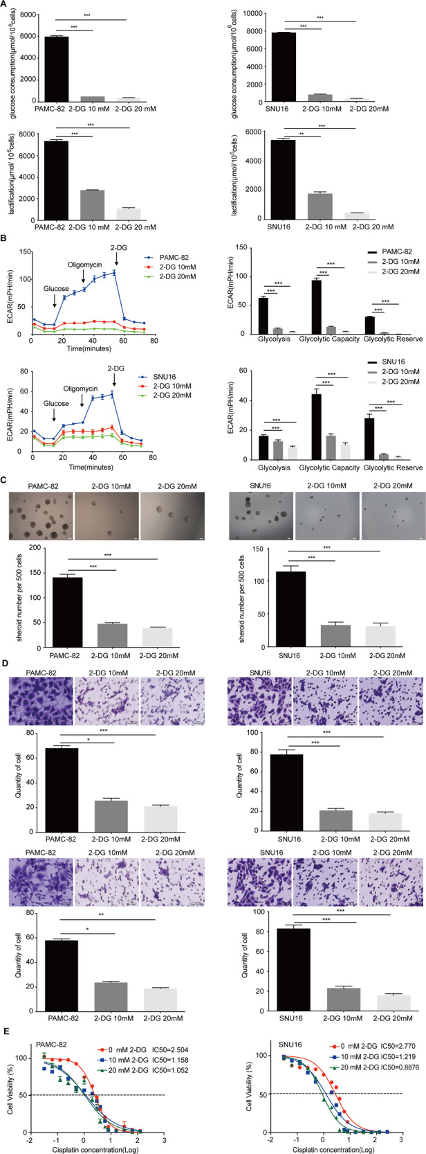
A PAMC-82 and SNU16 cells were cultured in 10 or 20 mM 2-DG for 36 h, and the levels of glucose consumption and lactic acid production were measured according to the cell numbers. Fold changes were then normalized (μmol/106 cells). B Extracellular acid ratio (ECAR) of cells was measured by Seahorse XF in PAMC-82 and SNU16 cells treated with 10 or 20 mM 2-DG for 36 h. ECAR curves of cells treated with glucose, oligomycin, or 2-DG. Black arrows indicate the time point of cell treatment. C Analysis of the self-renewal abilities of PAMC-82 and SNU16 cells cultured in 10 or 20 mM 2-DG for 24 h. Scale bar, 100 μm. D The migration and invasion assay of PAMC-82 and SNU16 cells treated with 10 or 20 mM 2-DG for 24 h (above: migration, below: invasion). Scale bar, 100 μm. E PAMC-82 and SNU16 cells cultured in 10 or 20 mM 2-DG for 24 h, in the presence of several different concentrations of cisplatin (0.015625–256 μM) for 72 h. Cell viability was measured by CCK8. Results are from representative experiments in triplicate and shown as the mean ± standard deviation (SD). *P < 0.05, **P < 0.01, ***P < 0.001.