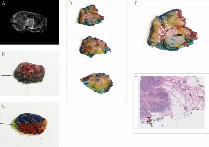
Figure 2.
Gross intraoperative assessment workflow with example specimen. (A) Radiograph of a freshly excised breast lumpectomy specimen. (B) Excised specimen with orienting sutures and a localization wire. Dye from the sentinel node localization procedure is present. (C) The specimen has been inked in six colors to designate the surgical margins (inferior, superior, anterior, posterior, medial, lateral). (D) Representative serial sections. A centrally located tumor is visible as a vaguely defined area of whitish discoloration. (E) A close-up with the region of tumor annotated (black circle), as well as the grossly identified closest margin (red ruler). In this case the green-inked inferior margin was closest, and grossly measured 1 mm to the tumor. A SAVI SCOUT localization device is present (Cianna Medical, Inc.). (F) Microscopic pathology showing invasive ductal carcinoma. The microscopic distance to the inferior margin was 1 mm (red ruler). Gross intraoperative assessment prompted immediate re-excision of this margin, and the re-excised margin was negative for carcinoma.

