Figure 6.

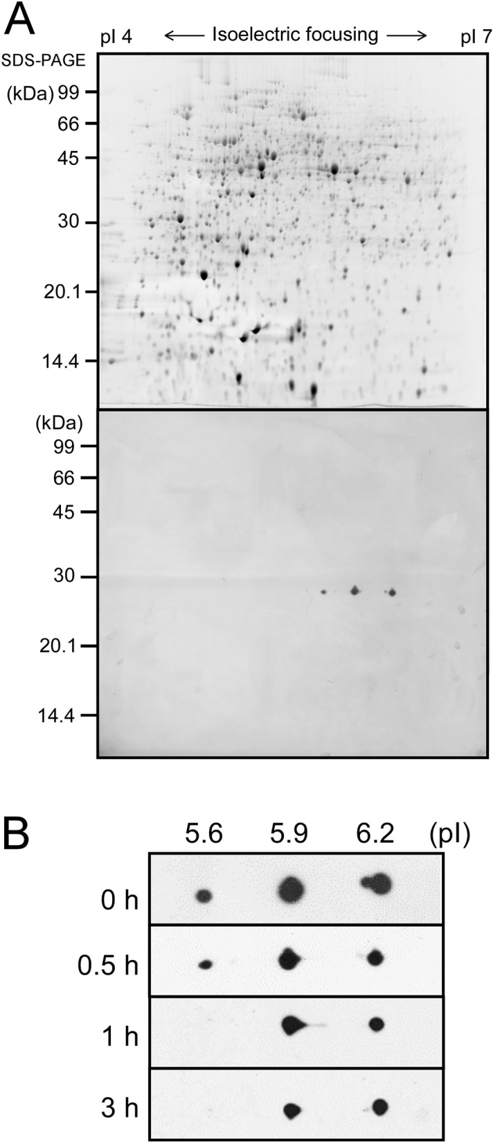
Changes in posttranslational modification of LexA upon the addition of 500 mM NaCl examined by 2D electrophoresis. (A) SYPRO Ruby-stained 2D gel with 270 μg of total protein from S.6803 grown under non-stress conditions (upper panel) and immunodetection of LexA using anti-LexA antibody (lower panel). At least three different forms of LexA with pIs of approximately 5.6, 5.9, and 6.2 were detected. (B) Changes in the spot pattern of LexA before and after the addition of NaCl at time points indicated.
