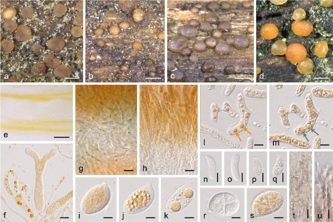
Fig. 5.

Pigments in the Dacrymycetes. a–d. Representative species with pulvinate and sessile (‘dacrymycetoid’) fruitbodies in the Dacryonaemataceae (a. Dacryonaema macnabbii, UPS F-940954), Unilacrymaceae (b. Unilacryma unispora, UPS F-941284), Cerinomycetaceae (c. Dacrymyces tortus s.lat. 3, UPS F-941018), Dacrymycetaceae (d. D. stillatus, UPS F-939816); e. yellow-orange spore print of Calocera viscosa (UPS F-940773); f, i–k. basidia and basidiospores of D. estonicus (UPS F-940137); g–h. reaction of the carotenoid contents with Lugol’s solution when applied from above to cuttings of D. estonicus (g, UPS F-940137) and U. unispora (h, UPS F-941284); l–m. dead arthrospores of D. stillatus (UPS F-941285) in water and after the application of Lugol’s solution, arrows depict the change in colour of lipid drops with carotenoids; n–o. basidiospores of Dacryonaema rufum (n, UPS F-941005) and Dacrymyces tortus s.lat. 4 (o, UPS F-941252); p–q. basidiospores of Calocera viscosa (UPS F-940773), before (p) and after (q) the coalescence of the lipid bodies; r–s. similarly shaped but differently pigmented basidiospores of Unilacryma unispora (r, UPS F-941280) and Dacrymyces ovisporus (s, UPS F-940139); t. cortical/marginal hyphae in Dacryonaema macrosporum (UPS F-941000), with brownish intracellular pigments; u. marginal hyphae in Da. rufum (UPS F-941005) with diffuse, brownish parietal pigment. — Scale bars: a–e = 1 mm, f–h = 10 μm, i–u = 5 μm.
