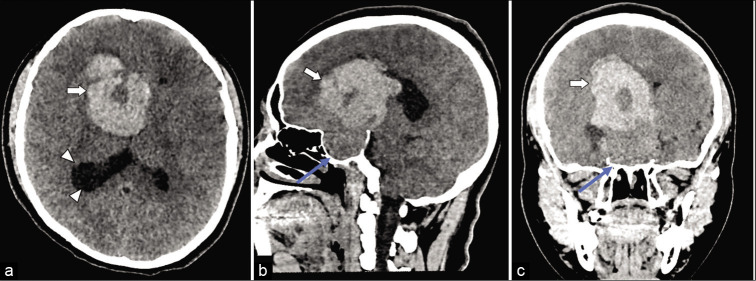
Figure 1:
Simple head CT scan obtained from axial plane (a), sagittal plane (b), and coronal plane (c). Heterogeneous tumor lesion of the sellar region is observed, with maximum dimensions of 59 × 52 × 68 mm (white arrow), causing widening of the sella turcica (blue arrow), with a hyperdense area compatible with tumor hemorrhage, causing collapse of the third ventricle and dilation of the occipital horn of the right lateral ventricle (arrowheads).

