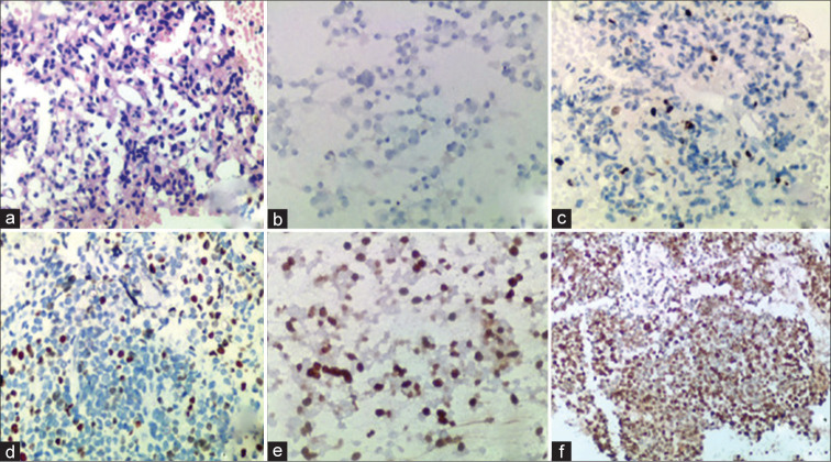
Figure 2:
(a) Cell block section of pancreatic neuroendocrine tumor (Pan NET) derived from EUS-FNA material (hematoxylin and eosin stain, ×20) (b) Pan NET G1 with Ki-67 positivity <3%, cell block (c) G2 with Ki-67 positivity between 3-20%, cell block (d) G3 WD Pan NET with Ki-67 positivity >20%, cell block (e) G3 WD Pan NET with Ki-67 positivity >20%, smear and (f) G3 PD Pan NEC with Ki-67 positivity >20%, cell block.

