
FIGURE 1.
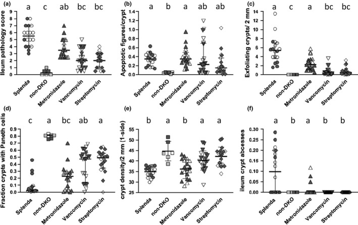
Ileum pathology scores and pathology marker levels in antibiotic‐treated DKO mice, Splenda‐treated DKO mice, and non‐DKO mice. Panel (A) is the overall score derived by adding values from appraisals of apoptosis (apoptosis figures/crypt >0.1 = 1; less = 0), Paneth cells (0.8–1 fraction crypts with Paneth cells = 0; 0–0.79 = 0.5; 0.34–0.59 = 1; 0.15–0.33 = 1.5; 0–0.14 = 2), crypt density (crypts/10× field one side; ≥39.1 = 0; 33.6–39 = 1; 27.8–33.5 = 2), inflammation foci/10× field both sides(0 = 0; 0.01–0.2 = 1; 0.21–0.5 = 2), and inflammation intensity (peak damage in section‐small abscesses = 1; large abscesses = 2; epithelial erosion/ulceration = 3; latter rarely found in B6); median and interquartile range. Panel B shows levels of crypt apoptosis, mean, and standard deviation. Panel (C) shows the fraction of crypts with anoikis (crypt exfoliation), median, and interquartile range. Panel (D) shows the fraction of crypts with Paneth cells, median and interquartile range. Panel (E) shows the number of crypt per 10× field, mean and standard deviation. Panel F shows the number of crypt abscesses per 10× field, median, and interquartile range. Data shown as median and interquartile range were evaluated by the Kruskal–Wallis test for multiple comparisons; data shown as mean and standard deviation were evaluated with 1‐way ANOVA with Tukey's multiple comparisons test. Common letters indicate no significant difference, otherwise a > b, etc. Open symbols for DKO sets (Splenda, metronidazole, vancomycin, and streptomycin) are males, dark symbols are females. For the non‐DKO mice, open symbols are Gpx1+/−Gpx2−/− males, dark symbols are Gpx1+/−Gpx2−/− females and half‐tone symbols are Gpx1−/−Gpx2+/− males