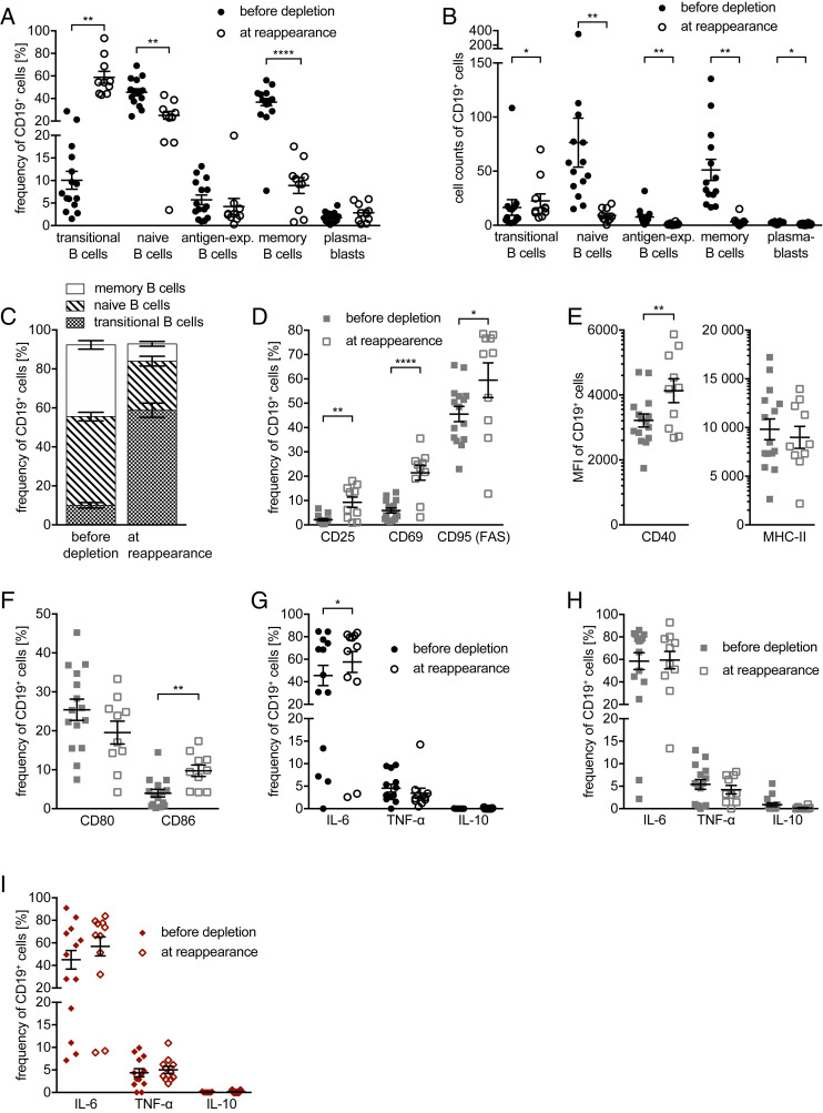
Fig. 3.
B cell maturation and phenotype before treatment initiation and at their reappearance. Peripheral blood mononuclear cells were isolated from 15 MS patients before anti-CD20 antibody treatment was initiated (filled shapes, before depletion; n = 15 samples) and after 8 to 24 mo (open shapes, at reappearance; n = 10 samples). Depicted are dot plots showing the mean ± SEM. Frequencies (A) and cell counts (B) of transitional (CD24high CD38high), mature naive (CD27− CD38+), antigen-experienced (=antigen-exp.; CD27+ CD38+), and memory (CD27var CD38−) B cells as well as plasmablasts (CD20− CD27+ CD38+) pre-gated on CD19+ B cells (*P < 0.05; **P < 0.005; ****P < 0.0001; Wilcoxon matched-pairs signed rank test/paired t test); frequency of transitional B cells and plasmablasts was gated within the mature naive respectively antigen-experienced B cells and was calculated to the B cell population. Second, frequency of transitional B cell and of plasmablasts was subtracted from mature naive respectively antigen-experienced B cells to receive the negative population. (C) Frequency of transitional, naïve, and memory B cells of all patients before depletion and at reappearance. Cells were cultured for 22 h in the presence of CpG (D–F; gray squares). Frequency (D) of CD19+ B cells expressing CD25, CD69, and CD95 (FAS) (*P < 0.05; **P < 0.005; ***P < 0.001; Wilcoxon matched-pairs signed rank test/paired t test). (E) B cells’ expression of CD40 and MHC class II (MHC-II) shown as mean fluorescence intensity (MFI) as well as (F) frequency of CD19+ B cells expressing CD80 and CD86 (**P < 0.005; Wilcoxon matched-pairs signed rank test/paired t test). Cells were cultured for 22 h unstimulated (G), in the presence of CpG (H), and 20 h unstimulated followed by 2 h in the presence of LPS (I), all followed by 4 additional hours in the presence of ionomycin, PMA, and GolgiPlug. Flow cytometry analysis of frequency of CD19+ B cells expressing tumor necrosis factor-α (TNF-α), interleukin-6 (IL-6), and IL-10 (*P < 0.05; Wilcoxon matched-pairs signed rank test).

