Fig. 2.
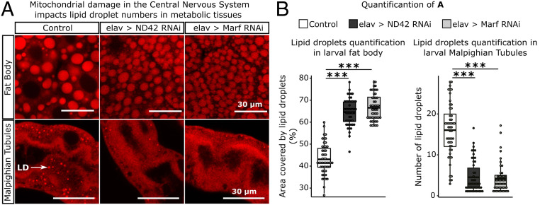
Inducing ROS in the brain impacts the lipid environment of metabolic tissues. (A) Knockdown of genes encoding mitochondrial components, ND42 and Marf, in neurons caused the accumulation of LDs in larval fat body and a reduction of LD numbers in larval Malpighian tubules. White arrow indicates LD in Malpighian tubules. Tissues were stained with Nile Red and images obtained in a Leica SP8 laser scanning confocal microscope, 400× magnification. Images were analyzed using the software ImageJ. (B) Quantification of the area covered by LDs per section of 50 µm × 50 µm in larval fat body; and number of LDs per section of 30 µm × 30 µm in larval Malpighian tubules (n = 10 larvae/treatment; 5 sections/larva). t test; ***P < 0.001.

