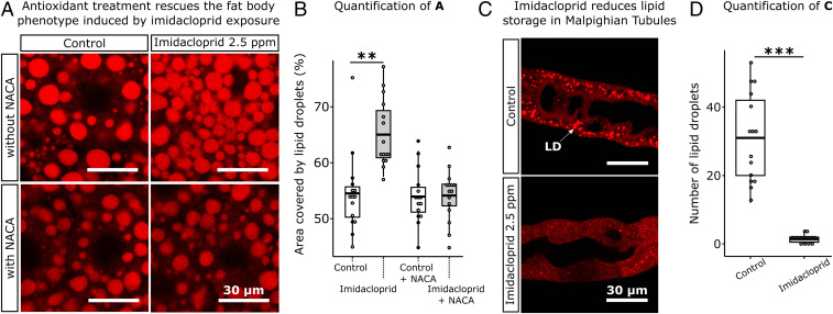
Fig. 3.
Imidacloprid perturbs the lipid environment of metabolic tissues and antioxidant treatment rescues the fat body phenotype. (A) Larvae exposed to imidacloprid show a significant increase in the area occupied by LDs in fat body, whereas larvae pretreated with 75 µg/mL of antioxidant NACA for 5 h prior to imidacloprid exposure show no significant changes in the area occupied by LDs in fat body. (Scale bar: 30 µm.) (B) Percentage of area occupied by LDs in fat body sections of 50 µm × 50 µm (n = 3 larvae/treatment; 5 sections/larva). (C) Reduced number of LDs in Malpighian tubules of larvae exposed to imidacloprid. White arrow indicates a LD. (D) Number of LDs per section of 30 µm × 30 µm in larval Malpighian tubules section of 30 µm × 30 µm (n = 3 larvae/treatment; 5 sections/larva). Images obtained in a Leica SP5 laser scanning confocal microscope, 400× magnification. t test; **P < 0.01; ***P < 0.001.

